. 2025 Jul 31;15(15):1932.
doi: 10.3390/diagnostics15151932.

Immunohistochemical and Ultrastructural Study of the Degenerative Processes of the Hip Joint Capsule and Acetabular Labrum

Affiliations

Immunohistochemical and Ultrastructural Study of the Degenerative Processes of the Hip Joint Capsule and Acetabular Labrum

Riana Maria Huzum et al. Diagnostics (Basel). .

Abstract

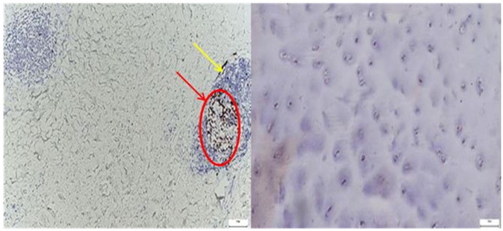
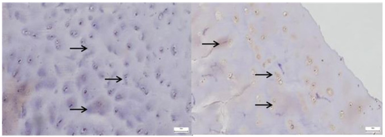
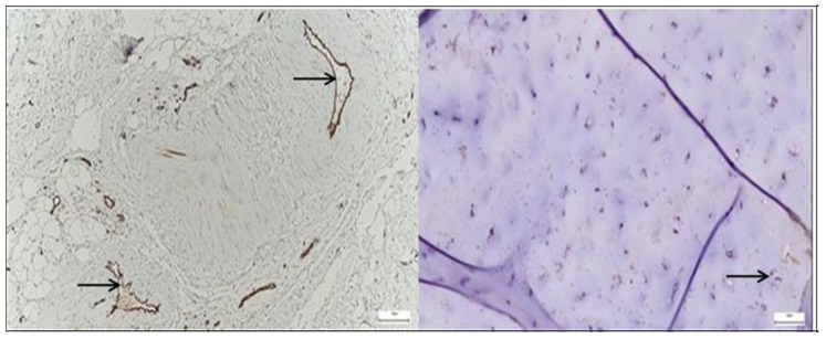
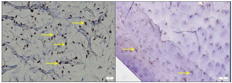
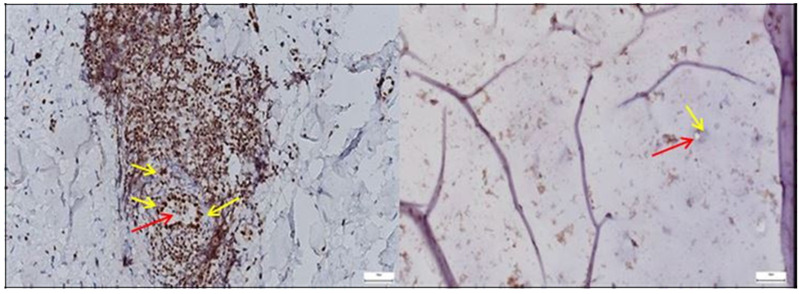
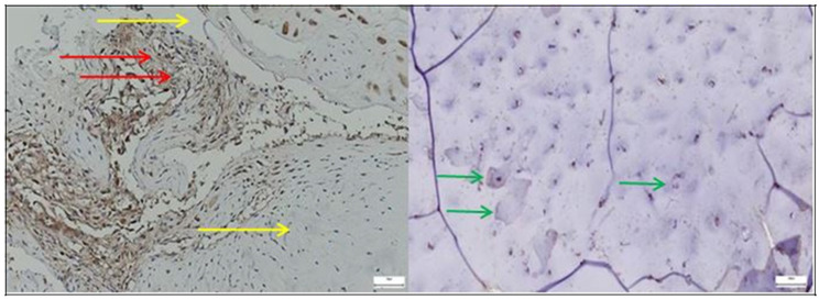
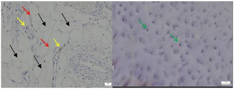
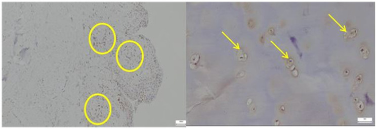
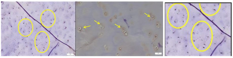
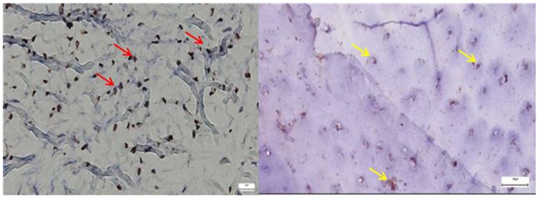
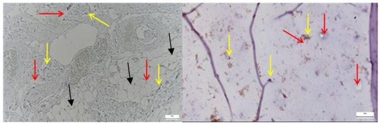
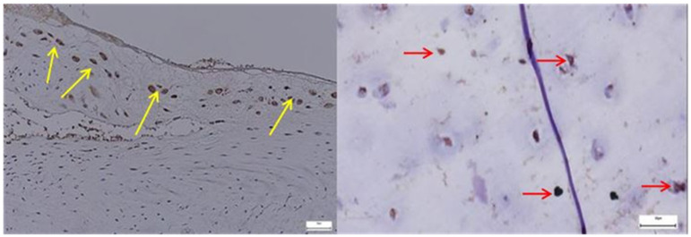
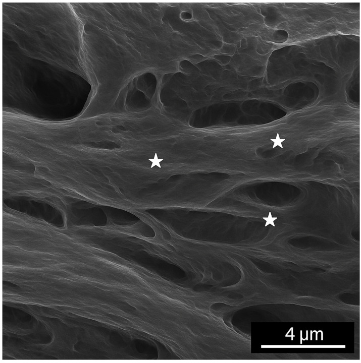
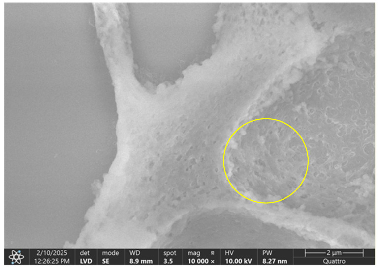
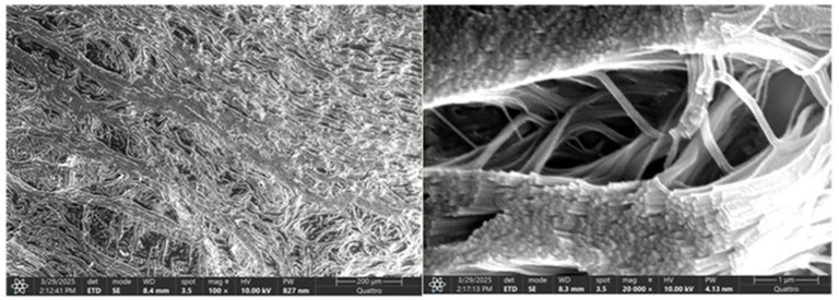
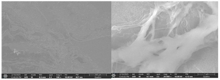
Background/Objectives: Degenerative processes of the hip joint increasingly affect not only the articular cartilage but also periarticular structures such as the joint capsule and acetabular labrum. This study aimed to investigate the structural and molecular changes occurring in these tissues during advanced hip osteoarthritis. Methods: A combined analysis using immunohistochemistry (IHC), scanning electron microscopy (SEM), and micro-computed tomography (microCT) was conducted on tissue samples from patients undergoing total hip arthroplasty and from controls with morphologically normal joints. Markers associated with proliferation (Ki67), inflammation (CD68), angiogenesis (CD31, ERG), chondrogenesis (SOX9), and lubrication (Lubricin) were evaluated. Results: The pathological group showed increased expression of Ki67, CD68, CD31, ERG, and SOX9, with a notable decrease in Lubricin. SEM analysis revealed ultrastructural disorganization, collagen fragmentation, and neovascular remodeling in degenerative samples. A significant correlation between structural damage and molecular expression was identified. Conclusions: These results suggest that joint capsule and acetabular labrum degeneration are interconnected and reflect a broader pathophysiological continuum, supporting the use of integrated IHC and SEM profiling for early detection and targeted intervention in hip joint disease.

Keywords: CD68; ERG; SEM; SOX9; acetabular labrum; hip capsule; immunohistochemistry; joint degeneration; lubricin; osteoarthritis.


Conflict of interest statement

The authors declare no conflicts of interest.

Figures

Figure 1
Figure 1
Schematic representation of the hip joint capsule and acetabular labrum anatomy in an oblique coronal section. The illustration highlights the anterosuperior quadrant of the acetabulum, showing the spatial relationship between the fibrous joint capsule and the fibrocartilaginous labrum. This orientation was chosen to emphasize the typical area affected by early degenerative changes, relevant for both arthroscopic access and pathological interpretation.
Figure 2
Figure 2
Representative anteroposterior pelvic radiographs illustrating the Kellgren–Lawrence grading system for hip osteoarthritis (Grades 0 to 4). Grade 0: Normal joint with preserved joint space and no osteophytes; Grade 1: Doubtful joint space narrowing and minimal osteophytic lipping; Grade 2: Definite osteophytes and possible joint space narrowing; Grade 3: Moderate osteophytes, definite joint space narrowing, and beginning subchondral sclerosis; Grade 4: Large osteophytes, severe joint space narrowing, pronounced subchondral sclerosis, and femoral head deformity. These radiographs were used as internal reference standards for classification in the pathological group.
Figure 3
Figure 3
Left—Immunohistochemistry for the cell proliferation marker Ki-67 in the hip joint capsule of a patient with degenerative joint disease. Perivascular lymphoid aggregates are seen, with a significant number of positively labeled nuclei (dark brown) in the germinal center of the follicles, indicating localized proliferative activity (red arrow and circle). The underlying tissue shows a reduced cell density and a disorganized collagen network, suggestive of chronic fibrous remodeling (yellow arrow). Magnification: 20×; scale: 50 µm; Right—Immunohistochemistry for Ki-67 in the control group. A homogeneous histological appearance is observed, with intact collagen organization and a reduced cell density, typical of healthy capsular tissue. Cell proliferation is minimal, being visible only by the rare presence of a few positively labeled nuclei (brown) in the stroma, without the formation of lymphoid aggregates or follicular proliferative activity. Magnification: 20×; scale: 50 µm.
Figure 4
Figure 4
Left—Immunohistochemistry for the CD68 marker in the hip joint capsule, taken from a patient with advanced degenerative pathology. Numerous positive cells (dark brown) diffusely dispersed in the stroma are highlighted (black arrows), with perivascular-subsynovial accumulations, corresponding to the infiltrate of activated macrophages. The presence of a small, poorly marked lymphoid aggregate suggests a nonspecific chronic inflammatory reaction. The appearance is suggestive of the involvement of monocyte/microphage components in tissue remodeling and the perpetuation of local chronic inflammation. Magnification: 20×; scale: 50 µm; Right—Immunohistochemistry for CD68 in normal hip joint capsule. A sparse distribution of positive cells (dark brown), with a predominantly perivascular location; resident population of tissue macrophages. The absence of diffuse infiltrates or lymphoid organization indicates a non-inflammatory tissue microenvironment, typical of a joint without active pathology. Magnification: 20×; scale: 50 µm.
Figure 5
Figure 5
Left—Immunohistochemistry for CD31 in the hip joint capsule, taken from a patient with degenerative pathology. An increased density of positive endothelial cells (dark brown) is highlighted, organized as mature vascular structures and neosynthesized capillaries, diffusely distributed in the stroma (black arrows). The appearance reflects an active process of neovascularization and microvascular remodeling, characteristic of chronic inflammation and progressive tissue degradation in degenerative arthropathy. Magnification: 20×; scale: 50 µm; Right—Immunohistochemistry for CD31 in a normal hip joint capsule. The presence of small, sparse vascular structures with weakly positive endothelial cells (brown), distributed peripherally and perivascularly, corresponding to a physiological capillary network, is observed. No areas of neovascularization or endothelial proliferation are evident, an appearance characteristic of capsular tissue without inflammation or active remodeling. Magnification: 20×; scale: 50 µm.
Figure 6
Figure 6
Left—In the pathological hip joint capsule, SOX9-positive nuclei appear more numerous and irregularly distributed within a disorganized fibrous matrix (yellow arrows), suggesting a reactive or compensatory upregulation of this transcription factor in response to structural and functional tissue alterations; Right—SOX9-positive nuclei are evenly distributed throughout the fibrous layer of the normal hip joint capsule, with stronger expression near fibrocartilaginous regions, reflecting its physiological role in maintaining matrix integrity and supporting joint tissue homeostasis, 40× bilateral.
Figure 7
Figure 7
Left—In the pathological hip joint capsule, ERG immunostaining (yellow arrows) revealed a heterogeneous pattern, with scattered nuclear positivity primarily confined to endothelial cells (red arrows) lining irregular microvascular structures, indicating altered angiogenic activity and potential vascular remodeling in the affected tissue; Right—In the normal hip joint capsule, ERG immunostaining showed discrete nuclear positivity restricted to endothelial cells of well-organized microvessels, consistent with physiological vascular architecture and baseline endothelial integrity, 25× bilateral.
Figure 8
Figure 8
Left—In the pathological hip joint capsule, Lubricin immunostaining revealed patchy and diminished expression (yellow arrows), with weak, or absent signal in areas of matrix disorganization (red arrows), suggesting impaired boundary lubrication and possible involvement in degenerative tissue changes. Right—In the normal hip joint capsule, Lubricin expression (green arrows) was clearly detected along the surface layers and within pericellular zones, with a continuous and well-defined immunostaining pattern, supporting its physiological role in joint lubrication and protection against mechanical stress, 40× bilateral.
Figure 9
Figure 9
Left—Ki67 immunohistochemical staining in the pathological acetabular labrum of the hip joint. The image shows scattered Ki67-positive nuclei (yellow arrows) within the fibrous and perivascular areas of the labral tissue, indicating localized proliferative activity. Increased nuclear labeling is observed particularly around vascular structures (red arrows) and within stromal regions, suggestive of ongoing remodeling or reparative responses associated with degenerative or inflammatory changes; black arrows mark the presence of adipose tissue. Right—Ki67 immunostaining in normal labral tissue. The image shows rare Ki67-positive nuclei within the fibrous matrix of the acetabular labrum, with most cells displaying no proliferative activity. The low density and scattered distribution of labeled nuclei are consistent with a quiescent cellular profile typical of healthy labral tissue under physiological conditions, 50× bilateral.
Figure 10
Figure 10
Left—CD68 immunostaining in pathological labral tissue. Numerous CD68-positive macrophages are evident (yellow circle), displaying a clustered and uneven distribution within the fibrous matrix and perivascular zones. This increased immunoreactivity reflects a prominent inflammatory response and suggests active monocyte-macrophage involvement in the pathological remodeling of the acetabular labrum; Right—CD68 immunostaining in normal labral tissue (yellow arrows), 40× bilateral.
Figure 11
Figure 11
Left—CD31 immunostaining in pathological labral tissue. The image reveals prominent CD31 expression in endothelial cells lining irregular and often enlarged microvascular profiles (yellow circle). The increased density and disorganized arrangement of CD31-positive vessels suggest active neoangiogenesis and vascular remodeling associated with pathological tissue changes; Midddle—CD31 immunostaining in normal labral tissue. Sparse CD31-positive endothelial cells are identified within well-formed microvascular structures (yellow arrows). The orderly distribution and low vessel density are consistent with the physiological vascular architecture of healthy labral tissue, lacking signs of angiogenic activation, 40× bilateral. The image on the right is a zoomed detail from the left panel, highlighting cell clusters and disrupted fiber arrangement in the degenerative capsule tissue (yellow circles).
Figure 12
Figure 12
Left—SOX9 immunostaining in pathological labral tissue (red arrows). The image displays an increased number of SOX9-positive nuclei, irregularly distributed within the fibrous matrix. The heightened and disorganized expression pattern suggests altered transcriptional regulation, possibly reflecting reactive or reparative responses to degenerative changes within the acetabular labrum; Right—SOX9 immunostaining in normal labral tissue (yellow arrows). Scattered SOX9-positive nuclei are observed within the fibrous component of the acetabular labrum, showing a low and uniform nuclear staining pattern. This limited expression is consistent with the physiological role of SOX9 in maintaining matrix integrity under non-pathological conditions, 25× bilateral.
Figure 13
Figure 13
Left—ERG immunostaining in pathological labral tissue. ERG-positive nuclei are prominently detected in endothelial cells lining irregular and densely packed microvessels (red arrows). The increased staining intensity and vascular complexity suggest endothelial activation and pathological neovascularization, consistent with vascular remodeling processes in degenerative labral tissue (yellow arrows); black arrows mark the presence of adipose tissue, with similar impact as in Figure 9; Right—ERG immunostaining in normal labral tissue. Sparse ERG-positive nuclei are observed within well-formed endothelial linings of small, regularly distributed microvessels. The low vascular density and orderly architecture are consistent with a quiescent endothelial phenotype typical of non-pathological labral tissue, 40× bilateral.
Figure 14
Figure 14
Left—Lubricin immunostaining in pathological labral tissue. The image shows a discontinuous and diminished Lubricin signal along the surface and pericellular regions of the acetabular labrum (yellow arrows). Fragmented and patchy expression patterns suggest compromised boundary lubrication and potential alterations in matrix integrity associated with degenerative changes; Right—Lubricin immunostaining in normal labral tissue (red arrows). A continuous and well-defined Lubricin signal is observed along the surface and pericellular regions of the acetabular labrum. This uniform expression pattern reflects intact boundary lubrication and matrix integrity, consistent with physiological homeostasis in healthy tissue, 40× bilateral.
Figure 15
Figure 15
Scanning electron microscopy (SEM) image of the normal hip joint capsule. White stars indicates connective tissue.
Figure 16
Figure 16
SEM image of the pathological hip joint capsule. The image illustrates a disrupted connective tissue structure, with partial loss of the typical stratified architecture. Collagen fibers appear fragmented and disorganized, lacking the parallel alignment seen in healthy tissue (yellow circle). The surface is irregular, with regions of matrix disintegration, indicating degenerative remodeling. These ultrastructural alterations suggest compromised mechanical integrity and tissue homeostasis.
Figure 17
Figure 17
Scanning electron microscopy (SEM) images of normal labral tissue—both panels illustrate the organized ultrastructural architecture of the healthy acetabular labrum. The surface appears smooth and continuous, with well-aligned collagen fibrils arranged in parallel bundles, indicative of mechanical integrity. No signs of surface disruption, fiber fragmentation, or matrix degradation are present, supporting the preservation of native extracellular matrix morphology under physiological conditions.
Figure 18
Figure 18
Scanning electron microscopy (SEM) images of pathological labral tissue—the images reveal marked ultrastructural disorganization of the labral matrix. Collagen fibers appear fragmented, irregularly oriented, and poorly compacted, with visible surface discontinuities and porous zones. These morphological alterations suggest matrix degradation and compromised mechanical stability, consistent with degenerative remodeling processes in the pathological acetabular labrum.

Similar articles

References

    1. Porta M., Pau M., Leban B., Deidda M., Sorrentino M., Arippa F., Marongiu G. Lower Limb Kinematics in Individuals with Hip Osteoarthritis during Gait: A Focus on Adaptative Strategies and Interlimb Symmetry. Bioengineering. 2021;8:47. doi: 10.3390/bioengineering8040047. - DOI - PMC - PubMed
    1. Steingrebe H., Spancken S., Sell S., Stein T. Effects of hip osteoarthritis on lower body joint kinematics during locomotion tasks: A systematic review and meta-analysis. Front. Sports Act. Living. 2023;5:1197883. doi: 10.3389/fspor.2023.1197883. - DOI - PMC - PubMed
    1. Toma A.G., Salahoru P., Hinganu M.V., Hinganu D., Cozma L.C.D., Patrascu A., Grigorescu C. Reducing the Duration and Improving Hospitalisation Time by Using New Surgical Tehniques and Psychotherapy. Rev. Chim. 2019;70:143–146. doi: 10.37358/RC.19.1.6869. - DOI
    1. Wang Z., Mao X., Guo Z., Zhao R., Feng T., Xiang C. Comparison of Walking Quality Variables between End-Stage Osteonecrosis of Femoral Head Patients and Healthy Subjects by a Footscan Plantar Pressure System. Medicina. 2022;59:59. doi: 10.3390/medicina59010059. - DOI - PMC - PubMed
    1. Nakajima A., Sakai R., Inoue E., Harigai M. Prevalence of patients with rheumatoid arthritis and age-stratified trends in clinical characteristics and treatment, based on the National Database of Health Insurance Claims and Specific Health Checkups of Japan. Int. J. Rheum. Dis. 2020;23:1676–1684. doi: 10.1111/1756-185X.13974. - DOI - PubMed

LinkOut - more resources