Review
. 2025 Aug 4;15(15):1953.
doi: 10.3390/diagnostics15151953.

Molar Pregnancy: Early Diagnosis, Clinical Management, and the Role of Referral Centers

Affiliations
Review

Molar Pregnancy: Early Diagnosis, Clinical Management, and the Role of Referral Centers

Antônio Braga et al. Diagnostics (Basel). .

Abstract

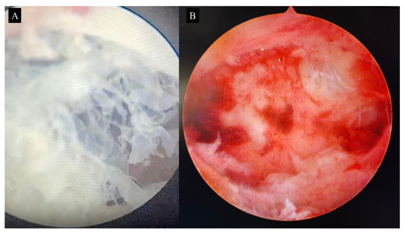
Molar pregnancy (MP) is a gestational disorder resulting from abnormal fertilization, leading to atypical trophoblastic proliferation and the formation of a complete or partial hydatidiform mole. This condition represents the most common form of gestational trophoblastic disease (GTD) and carries a significant risk of progression to gestational trophoblastic neoplasia (GTN). Although rare in high-income countries, MP remains up to ten times more prevalent in low-income and developing countries, contributing to preventable maternal morbidity and mortality. This narrative review provides an updated, practical overview of the clinical presentation, diagnosis, treatment, and follow-up of MP. A key focus is the challenge of early diagnosis, particularly given the increasing frequency of first-trimester detection, where classical histopathological criteria may be subtle, leading to diagnostic errors. The review innovates by integrating advanced diagnostic methods-combining histopathology, immunohistochemistry using p57Kip2, Ki-67, and p53 markers, along with cytogenetic analysis-to improve diagnostic accuracy in early gestation. The central role of referral centers is also emphasized, not only in facilitating timely treatment and access to chemotherapy, but also in implementing standardized post-molar follow-up protocols that reduce progression to GTN and maternal mortality. By focusing on both advanced diagnostic strategies and the organization of care through referral centers, this review offers a comprehensive, practice-oriented perspective to optimize patient outcomes in GTD and address persistent care gaps in high-burden regions.

Keywords: clinical presentation; diagnosis; follow-up; molar pregnancy; treatment.


Conflict of interest statement

The authors report no conflict of interest.

Figures

Figure 1
Figure 1
Macroscopic appearance of complete hydatidiform mole. Only the amorphous placental mass is visible, interspersed with translucent hydropic vesicles. Uterine evacuation was performed during the 8th week of gestation; thus, the vesicles are small, consistent with an early gestational-age mole. Note the absence of an embryo and embryonic appendages (such as yolk sac and umbilical cord).
Figure 2
Figure 2
Macroscopic appearance of partial hydatidiform mole. This molar pregnancy was interrupted at 14 weeks’ gestation. The placenta shows numerous hydropic vesicles scattered over the maternal surface.
Figure 3
Figure 3
Ultrasound of complete hydatidiform mole. The endometrial cavity is filled with an anechoic mass exhibiting multiple cystic areas (representing hydropic vesicles). No embryonic or appendageal structures are seen, but the uterus appears enlarged for gestational age, measuring 12 cm at its greatest diameter in what corresponds to an 8-week pregnancy.
Figure 4
Figure 4
Ultrasound of partial hydatidiform mole. A nonviable fetus at 14 weeks’ gestation is visible, along with a placental region containing two large anechoic areas.
Figure 5
Figure 5
Pathological findings of hydatidiform moles. (A) Representative case of complete hydatidiform mole (CHM). Note the presence of enlarged, hydropic chorionic villi with central cistern formation (black arrow), avascular stromal cores, and moderate circumferential trophoblastic hyperplasia (blue arrow) (Stain: Hematoxylin–Eosin). (B) Representative case of partial hydatidiform mole (PHM), showing a dual villous population composed of enlarged hydropic villi interspersed with smaller, more fibrotic ones. Finger-like projections (black arrow), trophoblastic invaginations (blue arrow), and intravillous inclusions (red arrow) are also observed (Stain: Hematoxylin–Eosin). (C) Absence of p57Kip2 immunostaining in both the villous cytotrophoblast (red arrow) and stromal cells (black arrow), consistent with the androgenetic origin and diagnosis of CHM. (D) Positive nuclear expression of p57Kip2 in cytotrophoblasts (red arrow) and villous stromal cells (black arrow), supporting the diagnosis of PHM. (E) Immunohistochemical staining for Ki-67 in CHM reveals a high proliferative index, with strong and widespread nuclear positivity in cytotrophoblastic and syncytiotrophoblastic cells, indicating intense mitotic activity. (F) In PHM, Ki-67 staining demonstrates a moderate proliferative index, with predominantly focal nuclear positivity in cytotrophoblasts and markedly lower labeling compared to CHM. (G) p53 immunostaining in CHM shows increased nuclear expression, particularly in cytotrophoblasts (semiquantitative score: 3+), with a diffuse to moderate distribution, reflecting deregulated cell cycle control and reduced apoptotic susceptibility. (H) In PHM, p53 expression is weak and focal (semiquantitative score: 1+), mainly restricted to cytotrophoblastic cells, with minimal to absent staining in syncytiotrophoblasts, consistent with the lower proliferative potential of PHM.
Figure 6
Figure 6
Image of a patient with 17 weeks of amenorrhea and a uterine fundal height of 22 cm, referred to the Gestational Trophoblastic Disease Referral Center at the Maternity School of Federal University of Rio de Janeiro with a diagnosis of molar pregnancy. Upon admission, the patient presented with profuse vaginal bleeding and expulsion of vesicles—a pathognomonic sign of the disease. Note the significant diagnostic delay, which led to clinical complications and the need for multiple blood transfusions, characterizing an obstetric near miss.
Figure 7
Figure 7
Image of a molar uterus. Hysterectomy was performed in a patient diagnosed with molar pregnancy at 48 years of age. Given advanced maternal age and completed parity, hysterectomy was chosen to reduce the risk of postmolar trophoblastic neoplasia. However, during follow-up, hCG levels remained elevated, and chemotherapy was required to achieve remission. This case highlights the necessity of post-hysterectomy hormonal surveillance.
Figure 8
Figure 8
(A) Manual intrauterine aspiration syringes and various sizes of aspiration cannulas. This is the most widely used equipment in Brazil for vacuum aspiration in definitive treatment of molar pregnancy. (B) Syringe filled with molar material freshly aspirated from the uterine cavity.
Figure 9
Figure 9
Pelvic ultrasound used to guide uterine evacuation for the treatment of molar pregnancy. Yellow arrows indicate the uterine aspiration cannula suctioning molar tissue. Ultrasound guidance during evacuation reduces the risk of uterine perforation and minimizes residual trophoblastic tissue.
Figure 10
Figure 10
Surgical hysteroscopy showing hydropic vesicles within the endometrial cavity (A). Following routine uterine evacuation, a repeat evaluation confirms complete removal of all trophoblastic tissue, ensuring the success of the procedure (B).

Similar articles

References

    1. Soper J.T. Gestational Trophoblastic Disease: Current Evaluation and Management. Obstet. Gynecol. 2021;137:355–370. doi: 10.1097/AOG.0000000000004240. - DOI - PMC - PubMed
    1. Lok C., van Trommel N., Braicu E.I., Planchamp F., Berkowitz R., Seckl M., EOTTD-ESGO-GCIG-ISSTD Guideline Committee Practical Guidelines for the Treatment of Gestational Trophoblastic Disease: Collaboration of the European Organisation for the Treatment of Trophoblastic Disease (EOTTD)-European Society of Gynaecologic Oncology (ESGO)-Gynecologic Cancer InterGroup (GCIG)-International Society for the Study of Trophoblastic Diseases (ISSTD) J. Clin. Oncol. 2025;43:2119–2128. - PubMed
    1. Horowitz N.S., Eskander R.N., Adelman M.R., Burke W. Epidemiology, diagnosis, and treatment of gestational trophoblastic disease: A Society of Gynecologic Oncology evidenced-based review and recommendation. Gynecol. Oncol. 2021;163:605–613. doi: 10.1016/j.ygyno.2021.10.003. - DOI - PubMed
    1. Joneborg U. Epidemiology of Gestational Trophoblastic Disease. Hematol. Oncol. Clin. N. Am. 2024;38:1173–1190. doi: 10.1016/j.hoc.2024.07.003. - DOI - PubMed
    1. Kaur B. Pathology of Gestational Trophoblastic Disease (GTD) Hematol. Oncol. Clin. N. Am. 2024;38:1191–1217. doi: 10.1016/j.hoc.2024.08.017. - DOI - PubMed

LinkOut - more resources