

The Role of Imaging in Ventricular Tachycardia Ablation
- PMID: 40804937
- PMCID: PMC12346019
- DOI: 10.3390/diagnostics15151973
The Role of Imaging in Ventricular Tachycardia Ablation
Abstract
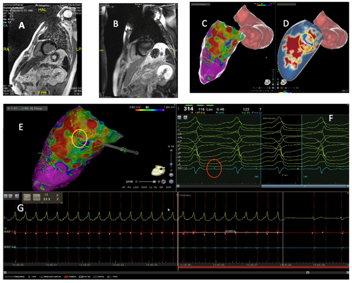
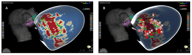
Ventricular tachycardia (VT) remains a major cause of morbidity and mortality in patients with structural heart disease. While catheter ablation has become a cornerstone in VT management, recurrence rates remain substantial due to limitations in electroanatomic mapping (EAM), particularly in cases of deep or heterogeneous arrhythmogenic substrates. Cardiac imaging, especially when multimodal and integrated with mapping systems, has emerged as a critical adjunct to enhance procedural efficacy, safety, and individualized strategy. This comprehensive review explores the evolving role of various imaging modalities, including echocardiography, cardiac magnetic resonance (CMR), computed tomography (CT), positron emission tomography (PET), and intracardiac echocardiography (ICE), in the preprocedural and intraprocedural phases of VT ablation. We highlight their respective strengths in substrate identification, anatomical delineation, and real-time guidance. While limitations persist, including costs, availability, artifacts in device carriers, and lack of standardization, future advances are likely to redefine procedural workflows.
Keywords: Imaging; cardiac magnetic resonance; catheter ablation; computed tomography; intracardiac echocardiography; ventricular tachycardia.
Conflict of interest statement
A.L.C. is employed by the company Johnson & Johnson Medial SpA. The remaining authors declare no conflicts of interest.
Figures
Similar articles
-
Comparing Low-to-Zero Fluoroscopic Navigation Systems for AVNRT Catheter Ablation: A Network Meta-Analysis.Pacing Clin Electrophysiol. 2024 Dec;47(12):1574-1585. doi: 10.1111/pace.15096. Epub 2024 Oct 22. Pacing Clin Electrophysiol. 2024. PMID: 39437197
-
Prescription of Controlled Substances: Benefits and Risks.2025 Jul 6. In: StatPearls [Internet]. Treasure Island (FL): StatPearls Publishing; 2025 Jan–. 2025 Jul 6. In: StatPearls [Internet]. Treasure Island (FL): StatPearls Publishing; 2025 Jan–. PMID: 30726003 Free Books & Documents.
-
Ablation of ventricular tachycardia from right ventricular aneurysms in patients with arrhythmogenic cardiomyopathy guided by intracardiac echocardiography.Heart Rhythm. 2025 Aug;22(8):1969-1974. doi: 10.1016/j.hrthm.2025.01.039. Epub 2025 Feb 3. Heart Rhythm. 2025. PMID: 39909314
-
Transfer of arrhythmia substrate targets from the cardiac electroanatomical and imaging modalities to the planning computed tomography scan for stereotactic arrhythmia radioablation for refractory ventricular tachycardia - a state-of-the-art review on software developments on behalf of the STOPSTORM.eu consortium.Radiother Oncol. 2025 Jun 27;210:111004. doi: 10.1016/j.radonc.2025.111004. Online ahead of print. Radiother Oncol. 2025. PMID: 40582572 Review.
-
Safety and efficacy of catheter ablation for ventricular tachycardia in elderly patients with structural heart disease: a systematic review and meta-analysis.J Interv Card Electrophysiol. 2023 Jan;66(1):179-192. doi: 10.1007/s10840-021-01007-w. Epub 2021 Aug 26. J Interv Card Electrophysiol. 2023. PMID: 34436722
References
-
- Zeppenfeld K., Tfelt-Hansen J., de Riva M., Winkel B.G., Behr E.R., Blom N.A., Charron P., Corrado D., Dagres N., de Chillou C., et al. 2022 ESC Guidelines for the management of patients with ventricular arrhythmias and the prevention of sudden cardiac death. Eur. Heart J. 2022;43:3997–4126. doi: 10.1093/eurheartj/ehac262. - DOI - PubMed
-
- Della Bella P., Baratto F., Vergara P., Bertocchi P., Santamaria M., Notarstefano P., Calò L., Orsida D., Tomasi L., Piacenti M., et al. Does Timing of Ventricular Tachycardia Ablation Affect Prognosis in Patients With an Implantable Cardioverter Defibrillator? Results From the Multicenter Randomized PARTITA Trial. Circulation. 2022;145:1829–1838. doi: 10.1161/CIRCULATIONAHA.122.059598. - DOI - PubMed
-
- Arenal Á., Ávila P., Jiménez-Candil J., Tercedor L., Calvo D., Arribas F., Fernández-Portales J., Merino J.L., Hernández-Madrid A., Fernández-Avilés F.J., et al. Substrate Ablation vs Antiarrhythmic Drug Therapy for Symptomatic Ventricular Tachycardia. J. Am. Coll. Cardiol. 2022;79:1441–1453. doi: 10.1016/j.jacc.2022.01.050. - DOI - PubMed
-
- Tung R., Xue Y., Chen M., Jiang C., Shatz D.Y., Besser S.A., Hu H., Chung F.-P., Nakahara S., Kim Y.-H., et al. First-Line Catheter Ablation of Monomorphic Ventricular Tachycardia in Cardiomyopathy Concurrent With Defibrillator Implantation: The PAUSE-SCD Randomized Trial. Circulation. 2022;145:1839–1849. doi: 10.1161/CIRCULATIONAHA.122.060039. - DOI - PubMed
Publication types
LinkOut - more resources
Full Text Sources
