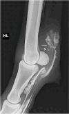
Does Low-Field MRI Tenography Improve the Detection of Naturally Occurring Manica Flexoria Tears in Horses?
- PMID: 40805040
- PMCID: PMC12345494
- DOI: 10.3390/ani15152250
Does Low-Field MRI Tenography Improve the Detection of Naturally Occurring Manica Flexoria Tears in Horses?
Abstract
Diagnosing digital flexor tendon sheath (DFTS) pathologies, particularly manica flexoria (MF) tears, can be challenging with standard imaging modalities. Standing low-field MRI tenography (MRIt) may improve the detection rate of MF tears. This study aimed to compare ultrasonography, contrast radiography, pre-contrast MRI, and MRIt to detect naturally occurring MF lesions in horses undergoing tenoscopy. Ten horses with a positive DFTS block, which underwent contrast radiography, ultrasonography, MRI, MRIt, and tenoscopy were included. Two radiologists evaluated the images and recorded whether an MF lesion was present and determined the lesion side. Sensitivity and specificity were calculated for each modality using tenoscopy as a reference. MRIt and contrast radiography detected MF lesions with the same frequency, both showing 71% sensitivity and 100% specificity. Pre-contrast MRI and ultrasonography detected MF lesions with a lower sensitivity (57%); however, the MRI (100%) demonstrated a higher specificity than ultrasonography (33%). Adding contrast in MRI changed the sensitivity from (4/7 lesions) 57% to (5/7 lesions) 71%, with a constant high specificity (100%). MRIt diagnoses MF tears with a similar sensitivity to contrast radiography, with the same specificity, but with the added benefit of lesion laterality detection. The combined advantages of the anatomical detail of the T1 sequence and the post-contrast hyperintense appearance of the fluid may help diagnose MF tears and identify intact MFs. However, this needs to be substantiated in a larger number of cases.
Keywords: MRI; horse; manica flexoria; tenoscopy.
Conflict of interest statement
The authors have no personal interests to declare.
Figures


References
-
- Arensburg L., Wilderjans H., Simon O., Dewulf J., Boussauw B. Nonseptic tenosynovitis of the digital flexor tendon sheath caused by longitudinal tears in the digital flexor tendons: A retrospective study of 135 tenoscopic procedures. Equine Vet. J. 2011;43:660–668. doi: 10.1111/j.2042-3306.2010.00341.x. - DOI - PubMed
-
- Wilderjans H., Boussau B., Madder K., Simon O. Tenosynovitis of the digital flexor tendon sheath and annular ligament constriction syndrome caused by longitudinal tears in the deep digital flexor tendon: A clinical and surgical report of 17 cases in Warmblood horses. Equine Vet. J. 2003;35:270–275. doi: 10.2746/042516403776148183. - DOI - PubMed
LinkOut - more resources
Full Text Sources

