Review
. 2025 Aug 4;15(15):2275.
doi: 10.3390/ani15152275.

Practical Review on Aetio-Pathogenesis and Symptoms in Pigs Affected by Clinical and Subclinical Oedema Disease and the Use of Commercial Vaccines Under Field Conditions

Affiliations
Review

Practical Review on Aetio-Pathogenesis and Symptoms in Pigs Affected by Clinical and Subclinical Oedema Disease and the Use of Commercial Vaccines Under Field Conditions

Juan Hernandez-Garcia et al. Animals (Basel). .

Abstract

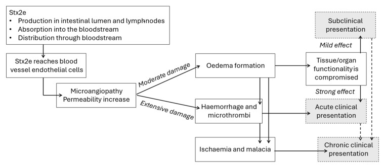
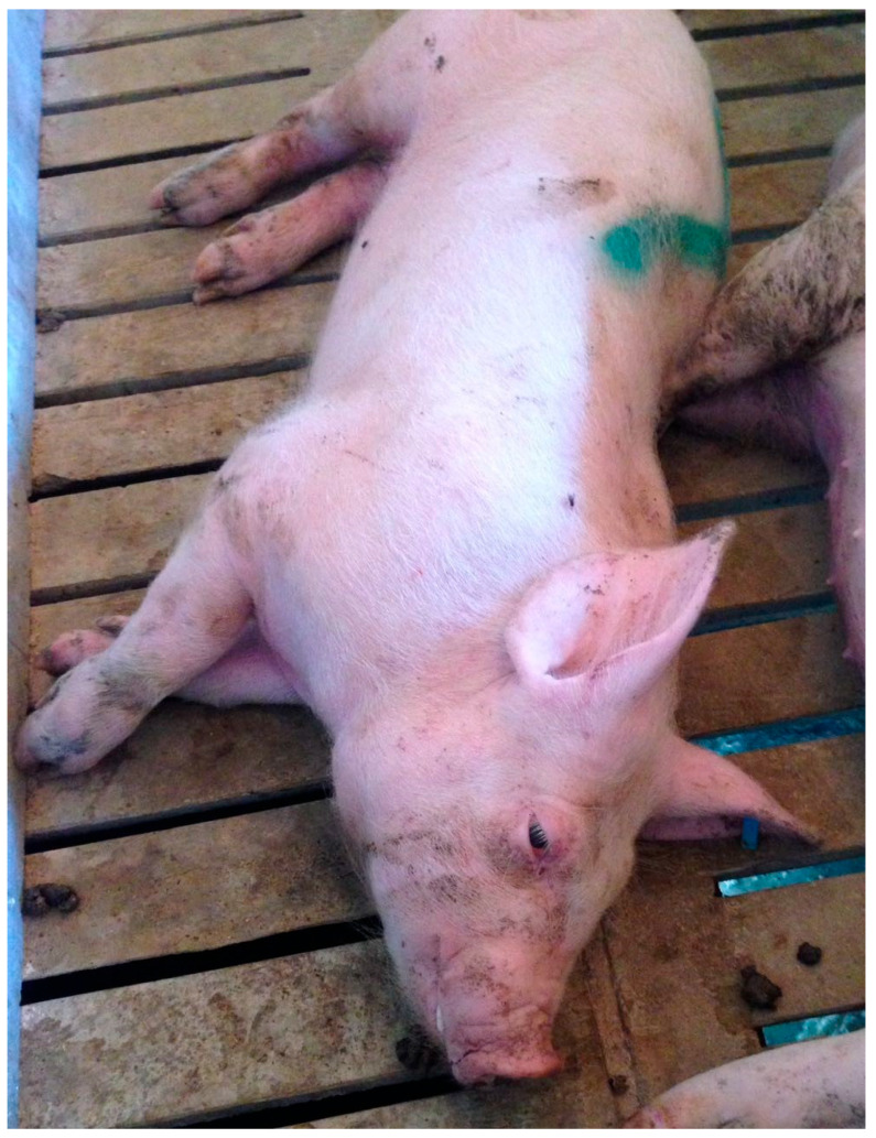
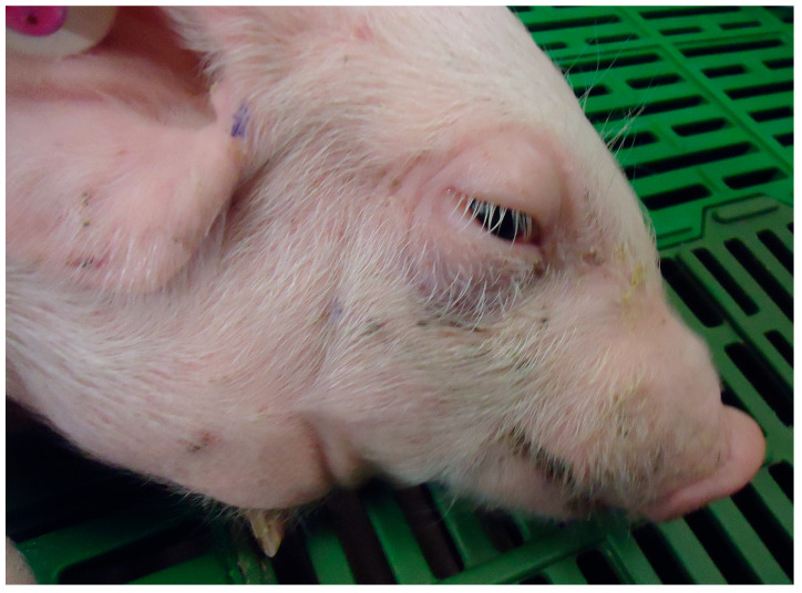
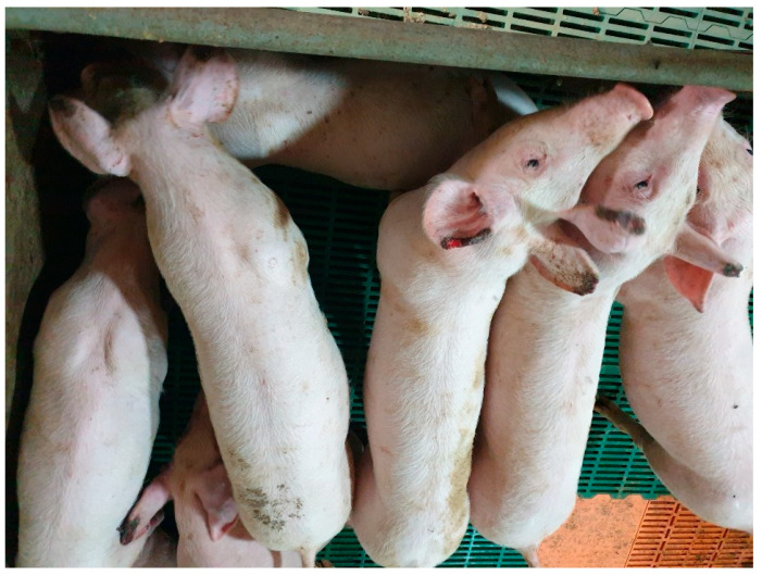
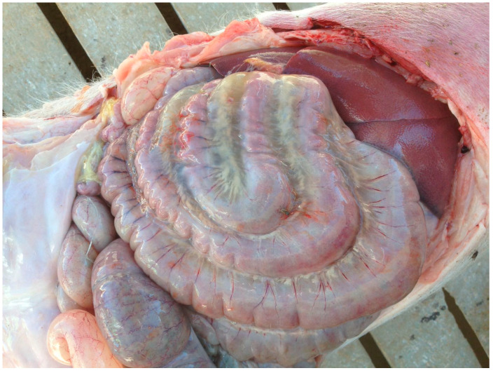
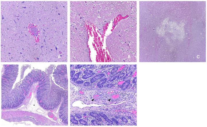
The impact of Oedema Disease produced by Shiga toxigenic Escherichia coli (STEC) in swine is increasing in some production countries due to increasing limitations on treatment with antimicrobials and zinc oxide, either because of the increased prevalence of multi-resistant strains or because of legal restrictions. The main pathological effect of Shiga toxin 2e is represented by damage to the endothelial cells of the blood vessel walls, leading to liquid extravasation and oedema formation in multiple tissues. These oedemas are generally easily identifiable in acute clinical cases. However, disease caused by Shiga toxin can occur without any externally visible oedema in the pigs, as observed in the subclinical presentation of Oedema Disease. It also causes productive losses, so it is important to identify and/or diagnose cases to set up control measures in order to optimize production and health. This article includes a comprehensive review of lesions and signs caused by Shiga toxin toxicosis in pigs, as well as other insights about the aetiology and epidemiology of STEC in pigs, and the effect of Shiga toxin recombinant toxoid vaccines in reducing these clinical and subclinical signs under field conditions.

Keywords: EDEC; STEC; Shiga toxigenic Escherichia coli; Shiga toxin 2e; Shiga toxin vaccine; Stx2e; edema disease Escherichia coli; oedema disease; oedema disease vaccines; recombinant Shiga toxin; subclinical oedema disease; verotoxin.


Conflict of interest statement

The authors declare that they work as the technical and marketing teams of HIPRA, which has a recombinant Shiga toxin Stx2e-based vaccine in its portfolio.

Figures

Figure 1
Figure 1
Toxin production and pathological effects producing different clinical presentations. Stx2e is produced by STEC in the intestinal lumen and/or lymph nodes, then absorbed into the bloodstream. Stx2e will produce its pathological effect on endothelial cells, causing increased microangiopathy and vessel permeability. Moderate pathological damage to vessels caused by Stx2e produces oedemas in surrounding tissues, which compromise tissue and organ function. Depending on the severity of this functionality loss, signs presented by affected animals are inapparent (or subclinical) when the effect is mild, or acute clinical when signs are evident. When Stx2e causes an extensive pathological damage, haemorrhages and microthrombi produce visible clinical signs (acute presentation). When lesions and poor tissue irrigation are maintained in time they can produce ischaemia and malacia. Oedemas, haemorrhages and microthrombi lesions can resolve and repair relatively fast and heal quickly, but once ischaemia and malacia lesions are produced, they will require more time if not chronic; a variable percentage of animals with clinical and sub-clinical signs do not successfully heal but develop chronic signs for a variable period of time.
Figure 2
Figure 2
Good quality pig affected by acute Oedema Disease; subcutaneous oedemas were visible in eyelids. Behavioural changes were severe; consciousness and reactivity to stimuli were reduced. Credits: Hipra archive.
Figure 3
Figure 3
Pig with Oedema Disease. Subcutaneous oedema is noticeable in eyelids, snout, forehead and neck. Credits: Hipra archive.
Figure 4
Figure 4
Ear necrosis cases observed in herd with an ongoing subclinical Oedema Disease problem. Credits: Hipra archive.
Figure 5
Figure 5
Gelatinous mesocolon oedema, which is considered a hallmark macroscopic lesion in fatalities caused by Oedema Disease. Credits: Hipra archive.
Figure 6
Figure 6
Histopathological images from a case of subclinical Oedema Disease [31]. (A) Thrombus and perivascular haemorrhage in the central nervous system (200× magnification); (B) Perivascular haemorrhage in the thalamus; (C) Focal spongiosis in the central nervous system (100× magnification). (D) Colonic submucosal oedema—lesion marked with asterisk—(100× magnification); (E) Fibrinoid degeneration, necrotizing vasculitis and thrombosis—lesion marked with arrow heads—in venules of colonic mucosa (200× magnification). Credits: UAB (Autonomous University of Barcelona) Veterinary Pathology Diagnostic Service.

References

    1. Fairbrother J.M., Nadeau É. Colibacillosis. In: Zimmerman J., Karriker L., Ramirez A., Schwartz K., Stevenson G., Zhang J., editors. Diseases of Swine. 11th ed. Wiley-Blackwell; Hoboken, NJ, USA: 2019. pp. 807–834. - DOI
    1. Wang X., Yu D., Chui L., Zhou T., Feng Y., Cao Y., Zhi S. A Comprehensive Review on Shiga Toxin Subtypes and Their Niche-Related Distribution Characteristics in Shiga-Toxin-Producing E. coli and Other Bacterial Hosts. Microorganisms. 2024;12:687. doi: 10.3390/microorganisms12040687. - DOI - PMC - PubMed
    1. Eriksen E.Ø., Pedersen K.S., Larsen I., Nielsen J.P. Evidence-Based Recommendations for Herd Health Management of Porcine Post-Weaning Diarrhea. Animals. 2022;12:1737. doi: 10.3390/ani12141737. - DOI - PMC - PubMed
    1. Canibe N., Højberg O., Kongsted H., Vodolazska D., Lauridsen C., Nielsen T.S., Schönherz A.A. Review on Preventive Measures to Reduce Post-Weaning Diarrhoea in Piglets. Animals. 2022;12:2585. doi: 10.3390/ani12192585. - DOI - PMC - PubMed
    1. Han C., Dai Y., Liu B., Wang L., Wang J., Zhang J. Diversity analysis of intestinal microflora between healthy and diarrheal neonatal piglets from the same litter in different regions. Anaerobe. 2019;55:136–141. doi: 10.1016/j.anaerobe.2018.12.001. - DOI - PubMed

LinkOut - more resources