Review
. 2025 Jul 31;17(15):2528.
doi: 10.3390/cancers17152528.

Neuroinflammation in Radiation Maculopathy: A Pathophysiologic and Imaging Perspective

Affiliations
Review

Neuroinflammation in Radiation Maculopathy: A Pathophysiologic and Imaging Perspective

Giulia Midena et al. Cancers (Basel). .

Abstract

Background: Radiation maculopathy (RM) is a delayed, sight-threatening complication of ocular radiotherapy. Traditionally regarded as a pure microvascular disease, emerging evidence points to the central role played by retinal neuroinflammation, driven by microglial activation and cytokine dysregulation affecting both the retina and the choroid. Hyperreflective retinal foci, neuroinflammatory in origin (I-HRF), visualized through advanced imaging modalities such as spectral domain optical coherence tomography (OCT), have been identified as early and critical biomarkers of both preclinical and clinical retinal neuroinflammation.

Materials and methods: This review synthesizes findings from experimental and clinical studies to explore the pathophysiology of neuroinflammation and the associated imaging parameters in RM.

Results: The integration of experimental and clinical evidence specifically underscores the significance of I-HRF as an early indicator of neuroinflammation in RM. OCT enables the identification and quantification of these biomarkers, which are linked to microglial activation and cytokine dysregulation.

Conclusions: The pathophysiology of RM has evolved from a predominantly vascular condition to one strongly secondary to neuroinflammatory mechanisms involving the retina and choroid. In particular, I-HRF, as early biomarkers, offers the potential for preclinical diagnosis and therapeutic intervention, paving the way for improved management of this sight-threatening complication.

Keywords: chorioretinal imaging; hyperreflective retinal foci; neuroinflammation; radiation maculopathy.


Conflict of interest statement

The authors declare no conflict of interest.

Figures

Figure 1
Figure 1
Color fundus photography of the left eye of a patient showing radiation maculopathy, characterized by ghost vessels (green arrows), hard exudates, and retinal hemorrhages. The subject was treated for a large-sized choroidal melanoma located in the inferotemporal quadrant. Plaque brachytherapy using Iodine-125 was performed, delivering a total dose of 85 Gy to the tumor apex. The image was acquired 25 months after radiation treatment. The patient had no history of diabetes mellitus.
Figure 2
Figure 2
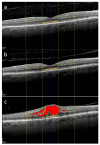
Overview of several spectral domain optical coherence tomography biomarkers evaluated in the macula of the right eye of a 62-year-old patient treated for uveal melanoma. The tumor was located in the inferior quadrant and classified as medium-sized. The patient underwent Iodine-125 plaque brachytherapy, with a total dose of 85 Gy delivered to the tumor apex. Intraretinal fluid (red); hyperreflective retinal foci (yellow dots) localized within the central 3 mm (yellow lines); external limiting membrane (orange) and ellipsoid zone (yellow) localized within the central 1 mm (green lines). 12 months after brachytherapy, hyperreflective retinal foci are 35, and they are mainly located in the inner retina (a). 24 months after treatment, there is an increase in the number of hyperreflective retinal foci with their progressive migration toward the outer retina (b). 30 months after brachytherapy, there is a further increase in the number of hyperreflective retinal foci with the appearance of center-involved macular edema (c). No treatment was administered between timepoints (b,c). Scale bar: 200 µm.
Figure 3
Figure 3
Identification of the subretinal fluid (blue) at spectral domain optical coherence tomography in the left eye of a 58-year-old patient treated for a large-sized choroidal melanoma located in the superior-nasal quadrant. The patient underwent Iodine-125 plaque brachytherapy with a total apex dose of 85 Gy. The subject had previously undergone a cycle of intravitreal anti-VEGF injections, which yielded no anatomical or functional improvement. Scale bar: 200 µm.
Figure 4
Figure 4
OCTA imaging of the superficial (a) and deep (b) capillary plexuses in the right eye of a 65-year-old patient treated for a large-sized choroidal melanoma located in the inferotemporal quadrant. The patient underwent Iodine-125 plaque brachytherapy with a total apex dose of 85 Gy. Both plexuses show capillary dropout and enlargement of the foveal avascular zone 20 months after treatment. No intravitreal treatment was administered prior to image acquisition. The patient had no history of diabetes mellitus.

Similar articles

  • Prescription of Controlled Substances: Benefits and Risks.
    Preuss CV, Kalava A, King KC. Preuss CV, et al. 2025 Jul 6. In: StatPearls [Internet]. Treasure Island (FL): StatPearls Publishing; 2025 Jan–. 2025 Jul 6. In: StatPearls [Internet]. Treasure Island (FL): StatPearls Publishing; 2025 Jan–. PMID: 30726003 Free Books & Documents.
  • Elbow Fractures Overview.
    Waseem M, Saeed W, Launico MV. Waseem M, et al. 2025 Jul 7. In: StatPearls [Internet]. Treasure Island (FL): StatPearls Publishing; 2025 Jan–. 2025 Jul 7. In: StatPearls [Internet]. Treasure Island (FL): StatPearls Publishing; 2025 Jan–. PMID: 28723005 Free Books & Documents.
  • Short-Term Memory Impairment.
    Cascella M, Al Khalili Y. Cascella M, et al. 2024 Jun 8. In: StatPearls [Internet]. Treasure Island (FL): StatPearls Publishing; 2025 Jan–. 2024 Jun 8. In: StatPearls [Internet]. Treasure Island (FL): StatPearls Publishing; 2025 Jan–. PMID: 31424720 Free Books & Documents.
  • Management of urinary stones by experts in stone disease (ESD 2025).
    Papatsoris A, Geavlete B, Radavoi GD, Alameedee M, Almusafer M, Ather MH, Budia A, Cumpanas AA, Kiremi MC, Dellis A, Elhowairis M, Galán-Llopis JA, Geavlete P, Guimerà Garcia J, Isern B, Jinga V, Lopez JM, Mainez JA, Mitsogiannis I, Mora Christian J, Moussa M, Multescu R, Oguz Acar Y, Petkova K, Piñero A, Popov E, Ramos Cebrian M, Rascu S, Siener R, Sountoulides P, Stamatelou K, Syed J, Trinchieri A. Papatsoris A, et al. Arch Ital Urol Androl. 2025 Jun 30;97(2):14085. doi: 10.4081/aiua.2025.14085. Epub 2025 Jun 30. Arch Ital Urol Androl. 2025. PMID: 40583613 Review.
  • Optic nerve head and fibre layer imaging for diagnosing glaucoma.
    Michelessi M, Lucenteforte E, Oddone F, Brazzelli M, Parravano M, Franchi S, Ng SM, Virgili G. Michelessi M, et al. Cochrane Database Syst Rev. 2015 Nov 30;2015(11):CD008803. doi: 10.1002/14651858.CD008803.pub2. Cochrane Database Syst Rev. 2015. PMID: 26618332 Free PMC article.

References

    1. Archer D.B., Amoaku W.M., Gardiner T.A. Radiation retinopathy—clinical, histopathological, ultrastructural and experimental correlations. Eye. 1991;5:239–251. doi: 10.1038/eye.1991.39. - DOI - PubMed
    1. Brown G.C., Shields J.A., Sanborn G., Augsburger J.J., Savino P.J., Schatz N.J. Radiation retinopathy. Ophthalmology. 1982;89:1494–1501. doi: 10.1016/S0161-6420(82)34611-4. - DOI - PubMed
    1. Midena G., Parrozzani R., Frizziero L., Midena E. Chorioretinal Side Effects of Therapeutic Ocular Irradiation: A Multimodal Imaging Approach. J. Clin. Med. 2020;9:3496. doi: 10.3390/jcm9113496. - DOI - PMC - PubMed
    1. Hawkins B.S. Collaborative Ocular Melanoma Study randomized trial of I-125 brachytherapy. Clin. Trials. 2011;8:661–673. doi: 10.1177/1740774511419684. - DOI - PubMed
    1. Melia B.M., Abramson D.H., Albert D.M., Boldt H.C., Earle J.D., Hanson W.F., Montague P., Moy C.S., Schachat A.P., Simpson E.R., et al. Collaborative ocular melanoma study (COMS) randomized trial of I-125 brachytherapy for medium choroidal melanoma I. visual acuity after 3 years COMS report no. 16. Ophthalmology. 2001;108:348–366. doi: 10.1016/s0161-6420(00)00526-1. - DOI - PubMed

LinkOut - more resources