. 2025 Jul 28;18(15):3538.
doi: 10.3390/ma18153538.

Long-Term Culture of Cellular Spheroids in Novel Hydrogel Constructs for ECM Characterization in Bone Models

Affiliations

Long-Term Culture of Cellular Spheroids in Novel Hydrogel Constructs for ECM Characterization in Bone Models

Diamante Boscaro et al. Materials (Basel). .

Abstract

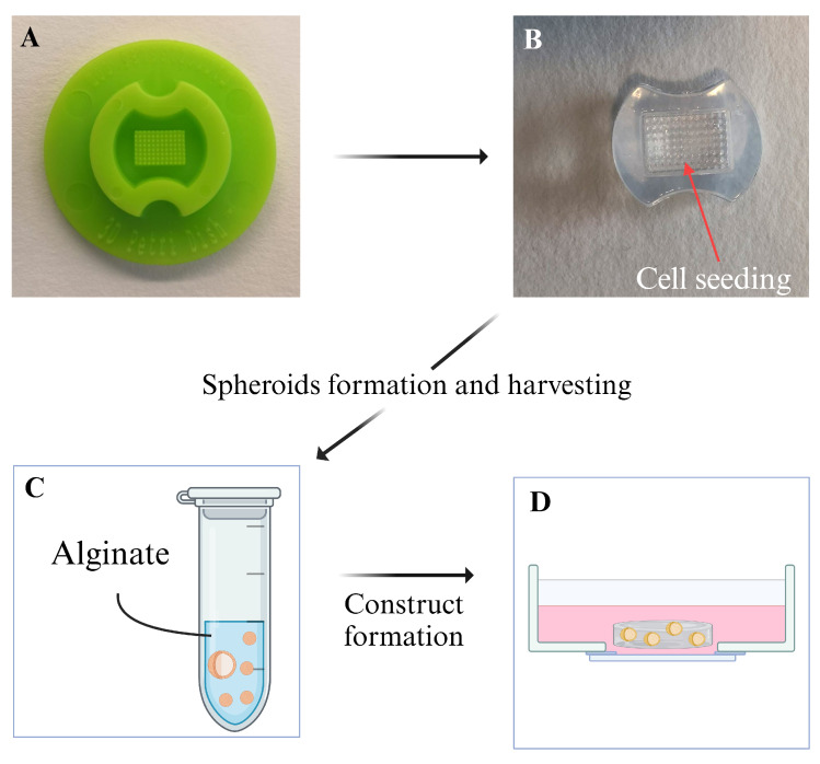
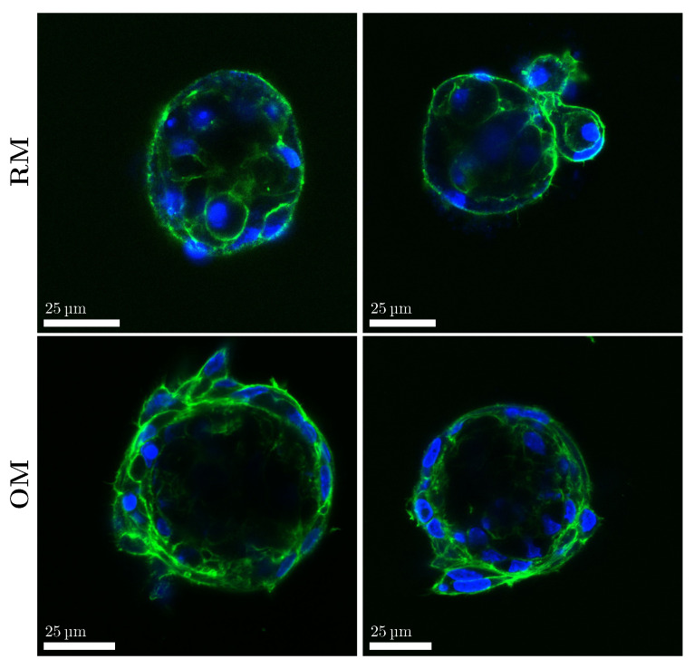
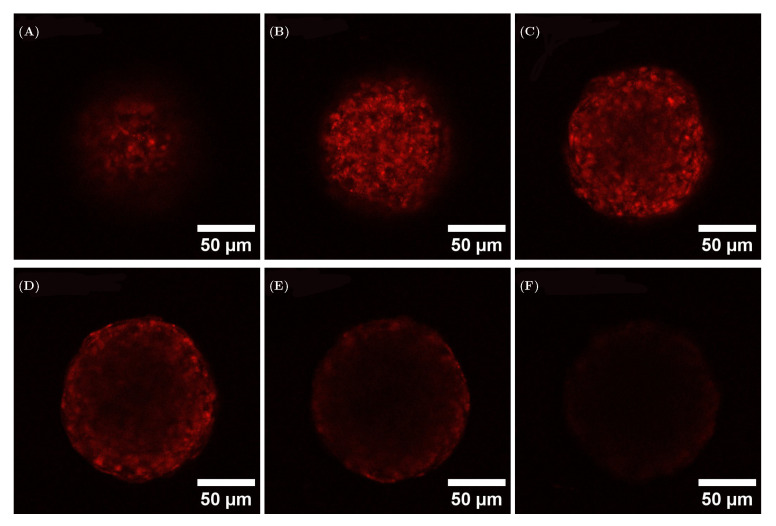
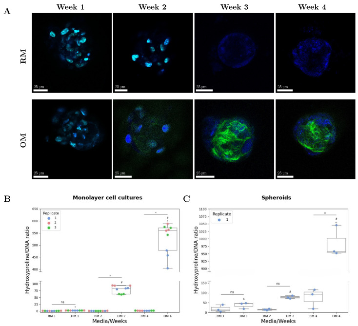
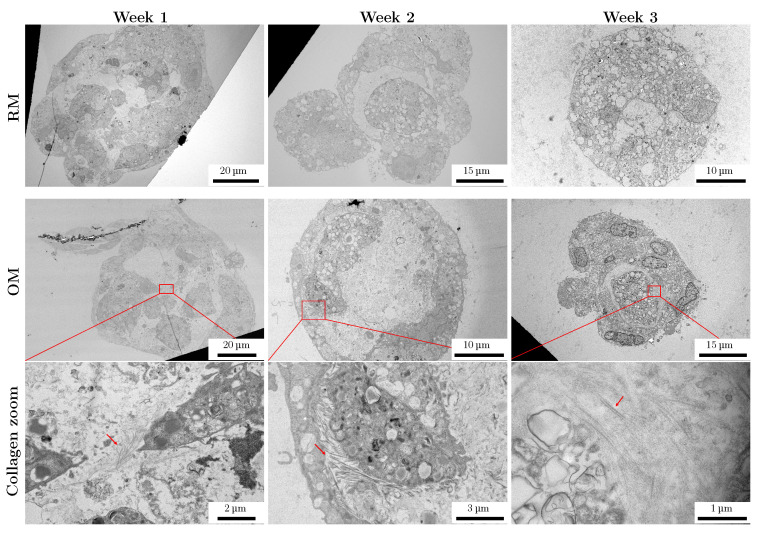
The application of cellular spheroids in bone tissue engineering research has gained significant interest in the last decade. Compared to monolayer cell cultures, the 3D architecture allows for more physiological cell-cell and cell-extracellular matrix (ECM) interactions that make cellular spheroids a suitable model system to investigate the bone ECM in vitro. The use of 3D model systems requires fine-tuning of the experimental methods used to study cell morphology, ECM deposition and mineralization, and cell-ECM interactions. In this study, we use a construct made of MC3T3-E1 cellular spheroids encapsulated in an alginate hydrogel to study and characterize the deposited ECM. Spheroid shape and structure were evaluated using confocal microscopy. The deposited collagenous ECM was characterized using Second Harmonic Imaging Microscopy (SHIM), quantitative hydroxyproline (HYP) assay, and Transmission Electron Microscopy (TEM). The use of hydrogel constructs enabled easy handling and imaging of the samples, while also helping to preserve the spheroid's stability by preventing cells from adhering to the culture dish surface. We used a non-modified alginate hydrogel that did not facilitate cell attachment and therefore functioned as an inert encapsulating scaffold. Constructs were cultured for up to 4 weeks. SHIM, HYP assay, and TEM confirmed the deposition of a collagenous matrix. We demonstrated that alginate-encapsulated bone spheroids are a convenient and promising model for studying the bone ECM in vitro.

Keywords: SHIM; TEM; collagen; immunofluorescence; spheroids.


Conflict of interest statement

The authors declare no conflicts of interest. The funders had no role in the design of the study; in the collection, analyses, or interpretation of data; in the writing of the manuscript; or in the decision to publish the results.

Figures

Figure 1
Figure 1
Schematic representation of spheroid formation. (A) The silicon mold used to create the (B) agarose molds. The cell suspension was placed in the central, pillared part of the mold. Spheroids were formed after ON incubation. (C) After collection from the molds, the spheroids were placed in a tube and resuspended in the alginate solution. (D) After gelation in the glass-bottomed Petri dish, the alginate disk containing the spheroids was kept in 2 mL of RM or OM.
Figure 2
Figure 2
Spheroids (gray spheres) in the mold, obtained after overnight incubation.
Figure 3
Figure 3
Spheroids showed a reduction in size during the first week of culture. (AF) Bright-field microscopy images of the spheroids encapsulated in the alginate hydrogel. The red arrows indicate the outline of the pocket that was formed due to the size reduction. (G) Quantification of the diameter reduction of spheroids cultured in RM and OM during the first week: spheroids were measured in the mold after overnight incubation (n = 97), after encapsulation (day 0) in RM (n = 64) and OM (n = 58), day 3 in RM (n = 73) and OM (n = 55), and day 7 in RM (n = 64) and OM (n = 96).
Figure 4
Figure 4
Shape analysis of spheroids cultured in RM and OM for 3 weeks. The upper images show bright-field images of the spheroids, and a difference in shape can be observed between the two different cultured samples. The lower images show immunofluorescence images of spheroids stained with nuclei (blue) and focal adhesion (red).
Figure 5
Figure 5
Cell morphology was influenced by the culturing conditions. Fluorescence images of actin (green) and cell nuclei (blue) of spheroids after 2 weeks in RM and OM.
Figure 6
Figure 6
Analysis of optical effects on spheroids, treated prior to aggregation with CellTracker Deep Red. The 6 cross-sections were taken with a step of 20 μm between each other, with the first section (A) starting at 5 μm. (B) 25 μm; (C): 45 μm; (D): 65 μm; (E): 85 μm; (F): 105 μm.
Figure 7
Figure 7
The osteogenic potential of alginate-encapsulated bone spheroids. (A) SHIM images of RM- and OM-cultured spheroids over 4 weeks. Collagen deposition (green) can be observed in OM-cultured spheroids at 2, 3, and 4 weeks. Spheroids cultured for 1 and 2 weeks were stained with Hoechst 34580 (blue), while spheroids cultured for 3 and 4 weeks were left unstained to ensure a better collagen signal. The blue signal observed in the unstained samples originated from 2-photon autofluorescence. Biochemical quantification of collagen in (B) monolayer cell cultures (n = 3) and (C) spheroids (n. of disks = 2). The HYP data were normalized to the DNA content. In spheroids, no statistical difference was observed between RM- and OM-cultured samples at week 1 (p = 0.979) and week 2 (p = 0.121), while a statistical difference was observed between RM- and OM-cultured samples at week 4 (p < 0.001). Significantly different data points are denoted with # and o for data points obtained at different time-points and with * when comparing data from the same time-point (RM v OM). Non-significant differences are noted with ns.
Figure 8
Figure 8
TEM images of 1-, 2-, and 3-week-old spheroids cultured in RM and OM. Regions of OM spheroids with collagen fibers are noted with a red rectangle, and a zoomed-in view of each region is shown below. Collagen fibers are indicated with red arrows.

References

    1. Agarwal R., García A.J. Biomaterial strategies for engineering implants for enhanced osseointegration and bone repair. Adv. Drug Deliv. Rev. 2015;94:53–62. doi: 10.1016/j.addr.2015.03.013. - DOI - PMC - PubMed
    1. Amini A.R., Laurencin C.T., Nukavarapu S.P. Bone Tissue Engineering: Recent Advances and Challenges. Crit. Rev. Biomed. Eng. 2012;40:363–408. doi: 10.1615/CritRevBiomedEng.v40.i5.10. - DOI - PMC - PubMed
    1. Urzì O., Gasparro R., Costanzo E., De Luca A., Giavaresi G., Fontana S., Alessandro R. Three-Dimensional Cell Cultures: The Bridge between In Vitro and In Vivo Models. Int. J. Mol. Sci. 2023;24:12046. doi: 10.3390/ijms241512046. - DOI - PMC - PubMed
    1. Huh D., Hamilton G.A., Ingber D.E. From 3D cell culture to organs-on-chips. Trends Cell Biol. 2011;21:745–754. doi: 10.1016/j.tcb.2011.09.005. - DOI - PMC - PubMed
    1. Liu S., Cheng L., Liu Y., Zhang H., Song Y., Park J.H., Dashnyam K., Lee J.H., Khalak F.A.H., Riester O., et al. 3D Bioprinting tissue analogs: Current development and translational implications. J. Tissue Eng. 2023;14:20417314231187113. doi: 10.1177/20417314231187113. - DOI - PMC - PubMed

LinkOut - more resources