

In Vitro Bioactivity and Cytotoxicity Assessment of Two Root Canal Sealers
- PMID: 40805596
- PMCID: PMC12348850
- DOI: 10.3390/ma18153717
In Vitro Bioactivity and Cytotoxicity Assessment of Two Root Canal Sealers
Abstract
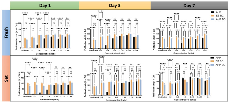
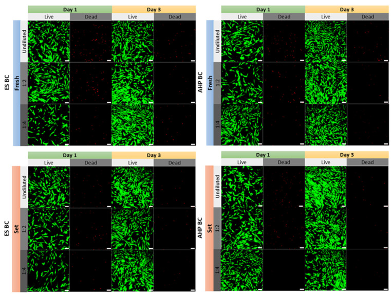
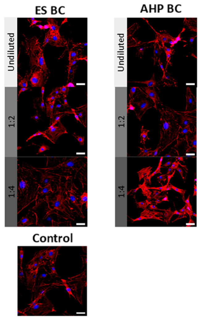
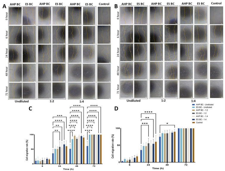
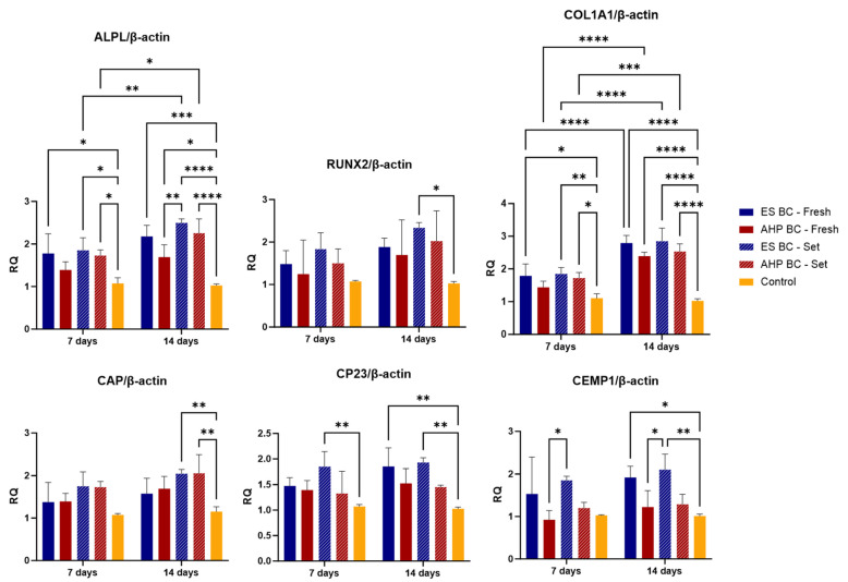
The development of bioactive materials in endodontics has advanced tissue regeneration by enhancing the biological responses of periradicular tissues. Recently, calcium silicate-based sealers have gained attention for their superior biological properties, including biocompatibility, osteoconductivity, and cementogenic potential. This study aimed to evaluate the cytotoxicity, biocompatibility, and bioactivity of EndoSequence BC Sealer (ES BC) and AH Plus Bioceramic Sealer (AHP BC) using human periodontal ligament stromal cells (hPDLSCs). Biocompatibility was assessed using MTT, Live/Dead, and wound healing assays. ES BC and AHP BC demonstrated significantly higher cell viability and proliferation compared to AH Plus used as a control. Gene expression analysis via real-time quantitative PCR demonstrated that ES BC, especially in set form, significantly upregulated osteogenic markers-alkaline phosphatase (2.49 ± 0.10, p < 0.01), runt-related transcription factor 2 (2.33 ± 0.13), and collagen type I alpha 1 chain (2.85 ± 0.40, p < 0.001)-more than cementogenic markers (cementum protein 1, cementum attachment protein, and cementum protein 23). This differential response may reflect the fibroblast-dominant nature of hPDLSCs, which contain limited cementoblast-like cells. This study supports the superior biocompatibility and regenerative capacity of ES BC and AHP BC compared to AH Plus. While in vitro models provide foundational insights, advanced ex vivo approaches are crucial for translating findings to clinical practice.
Keywords: PDL cells; bioceramic sealer; biocompatibility; cementogenesis; osteogenesis; periodontal ligament stem cells.
Conflict of interest statement
The authors declare no conflicts of interest.
Figures
Similar articles
-
From Cockle Shell to Bioceramic Root Canal Sealer: The Manufacturing and its Physical and Biological Properties.Int Dent J. 2025 Aug;75(4):100813. doi: 10.1016/j.identj.2025.03.022. Epub 2025 May 3. Int Dent J. 2025. PMID: 40318292 Free PMC article.
-
Cytocompatibility and bioactive potential of AH Plus Bioceramic Sealer: An in vitro study.Int Endod J. 2022 Oct;55(10):1066-1080. doi: 10.1111/iej.13805. Epub 2022 Aug 11. Int Endod J. 2022. PMID: 35950780 Free PMC article.
-
Biocompatibility and Bioactivity Evaluation of Novel Calcium Silicate-Based Sealer: In Vitro Study on Human Dental Pulp Stem Cells.Eur J Dent. 2025 Jul;19(3):777-783. doi: 10.1055/s-0045-1802566. Epub 2025 May 1. Eur J Dent. 2025. PMID: 40311629 Free PMC article.
-
Biological interactions between calcium silicate-based endodontic biomaterials and periodontal ligament stem cells: A systematic review of in vitro studies.Int Endod J. 2021 Nov;54(11):2025-2043. doi: 10.1111/iej.13600. Epub 2021 Aug 20. Int Endod J. 2021. PMID: 34338339
-
The efficacy of premixed bioceramic sealers versus standard sealers on root canal treatment outcome, extrusion rate and post-obturation pain: A systematic review and meta-analysis.Int Endod J. 2024 Aug;57(8):1021-1042. doi: 10.1111/iej.14069. Epub 2024 Apr 12. Int Endod J. 2024. PMID: 38606520
References
-
- Azim A.A., Aksel H., Zhuang T., Mashtare T., Babu J.P., Huang G.T.-J. Efficacy of 4 Irrigation Protocols in Killing Bacteria Colonized in Dentinal Tubules Examined by a Novel Confocal Laser Scanning Microscope Analysis. J. Endod. 2016;42:928–934. doi: 10.1016/j.joen.2016.03.009. - DOI - PMC - PubMed
LinkOut - more resources
Full Text Sources
Research Materials
