. 2025 Jul 31;14(15):2352.
doi: 10.3390/plants14152352.

Unlocking the Potential of Gracilaria chilensis Against Prostate Cancer

Affiliations

Unlocking the Potential of Gracilaria chilensis Against Prostate Cancer

Verónica Torres-Estay et al. Plants (Basel). .

Abstract

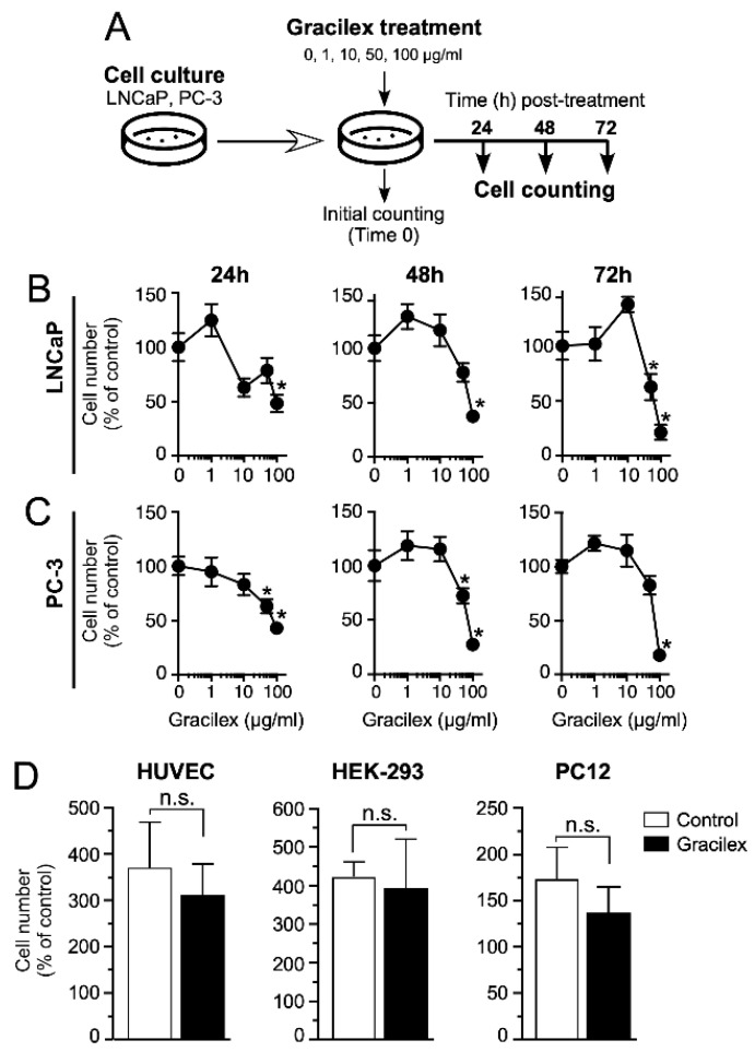
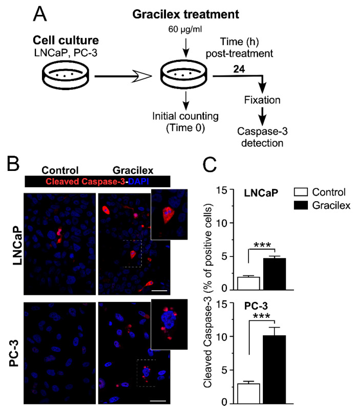
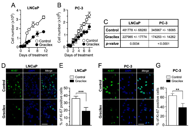
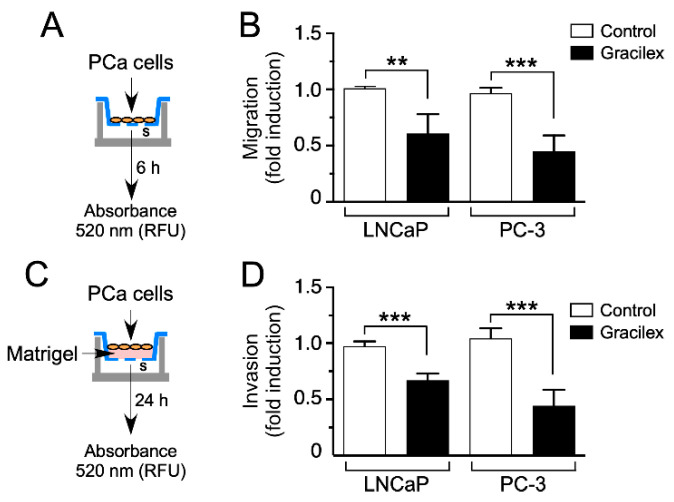
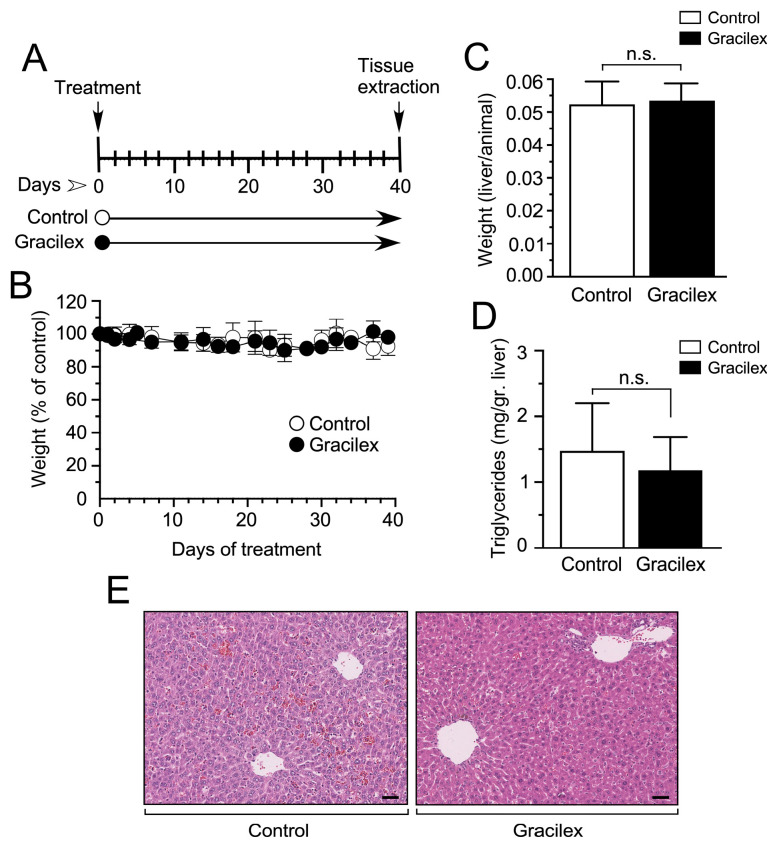
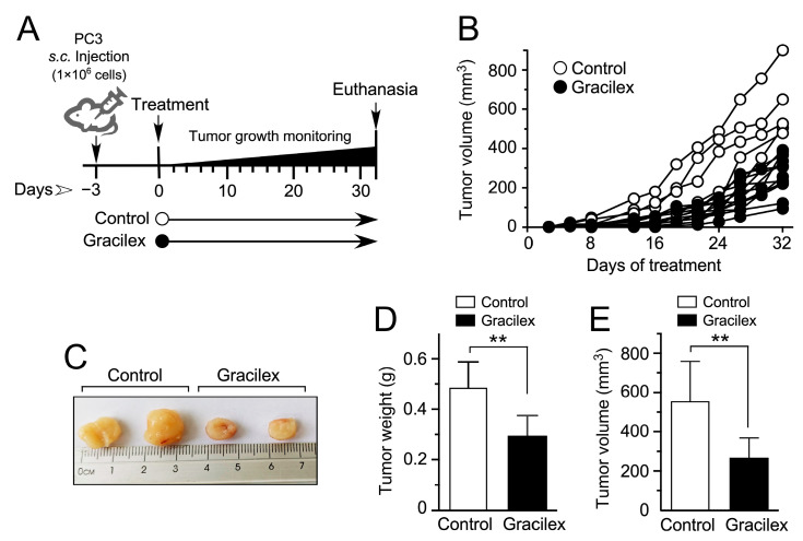
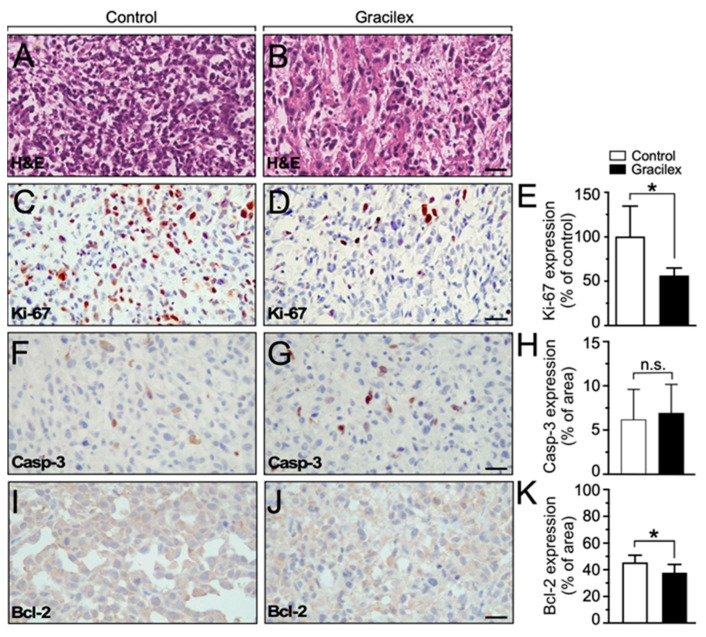
Prostate cancer (PCa) is the second leading cause of cancer-related death among men in most Western countries. Current therapies for PCa are limited, often ineffective, and associated with significant side effects. As a result, there is a growing interest in exploring new therapeutic agents, particularly from the polyphyletic group of algae, which offers a promising source of compounds with anticancer properties. Our research group has focused on investigating the effects of a novel oleoresin from Gracilaria chilensis, known as Gracilex®, as a potential therapeutic agent against PCa using both in vitro and in vivo models. Our findings indicate that Gracilex® exhibits a time- and dose-dependent inhibitory effect on cell survival in LNCaP and PC-3 PCa, reducing viability by over 50% and inducing apoptosis, as evidenced by a significant increase in activated caspase-3 expression in both cell lines. Moreover, Gracilex® significantly reduces the proliferation rate of both LNCaP and PC-3 prostate cancer cell lines, as evidenced by a marked decrease in the growth curve slope (p = 0.0034 for LNCaP; p < 0.0001 for PC-3) and a 40-50% reduction in the proportion of Ki-67-positive PCa cells. In addition, Gracilex® significantly reduces in vitro cell migration and invasion in LNCaP and PC-3 cell lines. Lastly, Gracilex® inhibits tumor growth in an in vivo xenograft model, an effect that correlates with the reduced PCa cell proliferation observed in tumor tissue sections. Collectively, our data strongly support the broad antitumoral effects of Gracilex® on PCa cells in vitro and in vivo. These findings advance our understanding of its potential therapeutic role in PCa and highlight the relevance of further investigating algae-derived compounds for cancer treatment.

Keywords: Gracilex®; cancer; oleoresin; prostate cancer; seaweed.


Conflict of interest statement

The authors declare no conflicts of interest.

Figures

Figure 1
Figure 1
Impact of Gracilex® on PCa cell viability. (A) Outline of the experimental procedure for treating and counting cells at 24, 48, and 72 h post-incubation with increasing concentrations of Gracilex® (0, 1, 10, 50, and 100 µg/mL). The 0 µg/mL concentration corresponded to the vehicle control condition (0.01% DMSO). (B,C) Cell counts for LNCaP and PC-3 cells at 24, 48, and 72 h following Gracilex® treatment. Data points represent the average of two independent experiments, each performed in triplicate. (D) Effect of Gracilex® on non-malignant cells and non-prostatic tumorigenic cell lines. HUVEC, HEK293, and PC12 cells were treated with Gracilex® (60 µg/mL), and cell counts were assessed at 48 h. Initial cell counts on day 0 were set as 100%. Statistical analysis was performed using a one-way ANOVA Test and a paired t-test. * p < 0.05. n.s.: not statistically significant.
Figure 2
Figure 2
Gracilex® induces apoptosis in PCa cells. (A) Schematic representation of the design for treating LNCaP and PC-3 cells with Gracilex® (60 µg/mL) and assessing the expression of the apoptotic marker-activated caspase-3 after 24 h. (B) Immunofluorescence staining of activated caspase-3 in LNCaP and PC-3 cells, visualized using Alexa Fluor 594-conjugated anti-rabbit IgG. Nuclei were counterstained with DAPI. Scale bars: 20 µm. (C) Quantification of activated caspase-3-positive cells in LNCaP and PC-3 cell lines, expressed as a percentage of total cells based on DAPI staining. Statistical analysis was performed using an unpaired t-test. *** p < 0.001.
Figure 3
Figure 3
Effect of Gracilex® on cell growth and proliferation in PCa cells. LNCaP (A) and PC-3 (B) cells were counted daily from day 0 to day 10 for vehicle (control) and Gracilex®-treated cells (60 µg/mL). Each data point represents the mean of three independent experiments performed in triplicate. (C) The table shows the changes in the slopes of each growth curve in the absence (control) and presence of Gracilex® (60 µg/mL) for LNCaP and PC-3 cell lines. (D) Immunofluorescence analysis of the proliferation marker Ki-67 in LNCaP cells exposed to vehicle (control) or Gracilex® (60 µg/mL). (E) Quantification of Ki-67-positive LNCaP cells, expressed as a percentage of the total cell population based on DAPI staining. (F) Immunofluorescence analysis of the proliferation marker Ki-67 in PC-3 cells exposed to vehicle (control) or Gracilex® (60 µg/mL). (G) Quantification of Ki-67-positive PC-3 cells, expressed as a percentage of the total cell population based on DAPI staining. Nuclei were counterstained with DAPI. White scale bars: 20 µm. Statistical analysis was performed using an unpaired t-test. ** p < 0.005, *** p < 0.001.
Figure 4
Figure 4
Effect of Gracilex® on migration and invasion capacities in PCa cells. (A,C) Schematic representations of the experimental design for treating LNCaP and PC-3 cells with Gracilex® (60 µg/mL) to assess their migration and invasion capacities. (B) Migration and (D) invasion capacities were measured using the CytoSelect™ Migration and Invasion Assay. Vehicle (control) or Gracilex® treatments were applied to both the upper and lower chambers. FBS (serum, s) was used as a chemoattractant in the lower chamber. For invasion assays, the upper chamber contained a layer of Matrigel (C). The number of migrating/invading cells was quantified in the lower chamber using a fluorescent dye and measured with a Synergy Plate Reader. Statistical analysis was performed using an unpaired t-test. ** p < 0.005, *** p < 0.001.
Figure 5
Figure 5
Effect of Gracilex® on in vivo mouse models. (A) Schematic representation of the experimental design for treating mice with either vehicle (control) or Gracilex® (300 mg/kg). (B) The effect of corn oil (control) or Gracilex® treatment on the total body weight of mice, measured every 2 days for 40 days. (C) The liver-to-body weight ratio was measured at the end of the treatment period (40 days). (D) Triglyceride levels per gram of liver tissue were measured at the end of the treatment period (40 days). (E) Hematoxylin and eosin staining of liver sections from mice treated with vehicle (control) or Gracilex®. Scale bars: 40 µm. Statistical analysis was performed using an unpaired t-test. n.s. = not statistically significant.
Figure 6
Figure 6
Effect of Gracilex® on a cell line-derived xenograft model of human PCa. (A) Schematic representation of the experimental design for treating mice bearing PC-3 cell line-derived tumors with either corn oil (control) or Gracilex® (300 mg/Kg) for 4–5 weeks. (B) Tumor growth of PC-3 cell line-derived xenografts was measured 2–4 times per week. (C) Representative images of dissected PC-3 cell line-derived tumors. (D,E) Tumor weight (g) and volume (mm3) were measured after dissecting tumors from mice treated with vehicle (control) or Gracilex® (60 µg/mL). Statistical analysis was performed using an unpaired t-test. ** p < 0.005.
Figure 7
Figure 7
Effect of Gracilex® on in vivo expression of cell proliferation and apoptotic markers. (A,B) Hematoxylin and eosin staining of tissue sections from cell line-derived xenograft tumors treated with either corn oil (control) or Gracilex® (300 mg/Kg). (C,D) Immunohistochemical analysis of Ki-67 in tissue sections from cell line-derived xenograft tumors treated with either corn oil (control) or Gracilex® (300 mg/Kg). (E) Quantification of Ki-67 immunostaining, expressed as the percentage of Ki-67-positive nuclei relative to the total cell population based on hematoxylin staining. (F,G) Immunohistochemical analysis of activated caspase-3 in tissue sections from cell line-derived xenograft tumors treated with either corn oil (control) or Gracilex® (300 mg/Kg). (H) Quantification of cleaved caspase-3 immunostaining, expressed as the percentage of the total area of the tissue section. (I,J) Immunohistochemical analysis of Bcl-2 in tissue sections from cell line-derived xenograft tumors treated with either corn oil (control) or Gracilex® (300 mg/Kg). (K) Quantification of Bcl-2 immunostaining, expressed as the percentage of the total area of the tissue section. Scale bars: 20 µm. Statistical analysis was performed using an unpaired t-test. * p < 0.05, n.s. = not statistically significant.

Similar articles

References

    1. Miller K.D., Nogueira L., Devasia T., Mariotto A.B., Yabroff K.R., Jemal A., Kramer J., Siegel R.L. Cancer treatment and survivorship statistics, 2022. CA Cancer J. Clin. 2022;72:409–436. doi: 10.3322/caac.21731. - DOI - PubMed
    1. Siegel R.L., Kratzer T.B., Giaquinto A.N., Sung H., Jemal A. Cancer statistics, 2025. CA Cancer J. Clin. 2025;75:10–45. doi: 10.3322/caac.21871. - DOI - PMC - PubMed
    1. Sung H., Ferlay J., Siegel R.L., Laversanne M., Soerjomataram I., Jemal A., Bray F. Global Cancer Statistics 2020: GLOBOCAN Estimates of Incidence and Mortality Worldwide for 36 Cancers in 185 Countries. CA Cancer J. Clin. 2021;71:209–249. doi: 10.3322/caac.21660. - DOI - PubMed
    1. Diaz K.A., Amaya S.L., Garcia-Perdomo H.A. Perspectives on prostate cancer: Advances and pending challenges for a multidisciplinary oncological approach in South America. Int. Urol. Nephrol. 2024;56:1–7. doi: 10.1007/s11255-023-03753-4. - DOI - PMC - PubMed
    1. Catalona W.J., Smith D.S., Ratliff T.L., Basler J.W. Detection of organ-confined prostate cancer is increased through prostate-specific antigen-based screening. JAMA. 1993;270:948–954. doi: 10.1001/jama.1993.03510080052031. - DOI - PubMed

LinkOut - more resources