. 2025 Aug 3;14(15):2399.
doi: 10.3390/plants14152399.

Identification and Characterization of NAC Transcription Factors Involved in Pine Wilt Nematode Resistance in Pinus massoniana

Affiliations

Identification and Characterization of NAC Transcription Factors Involved in Pine Wilt Nematode Resistance in Pinus massoniana

Zhengping Zhao et al. Plants (Basel). .

Abstract

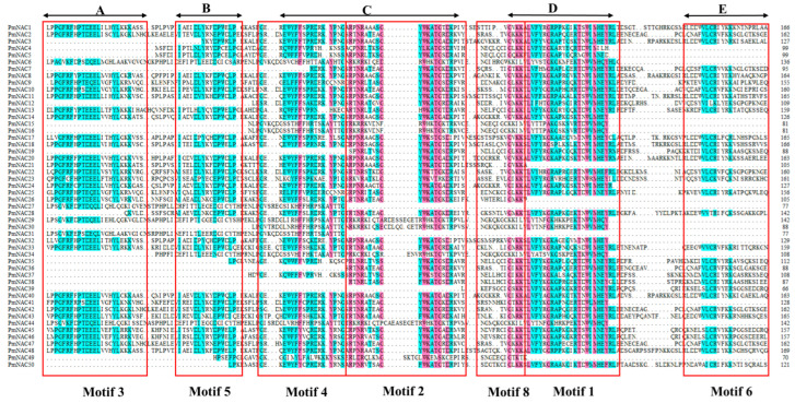
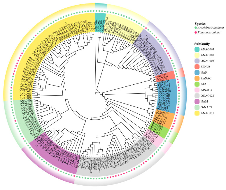
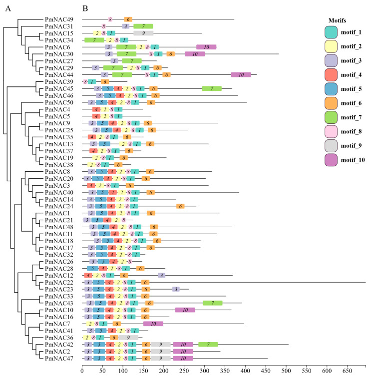
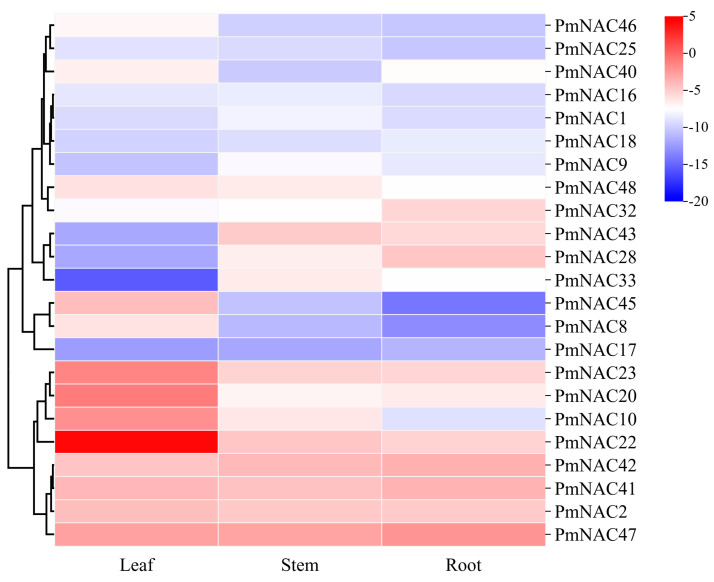
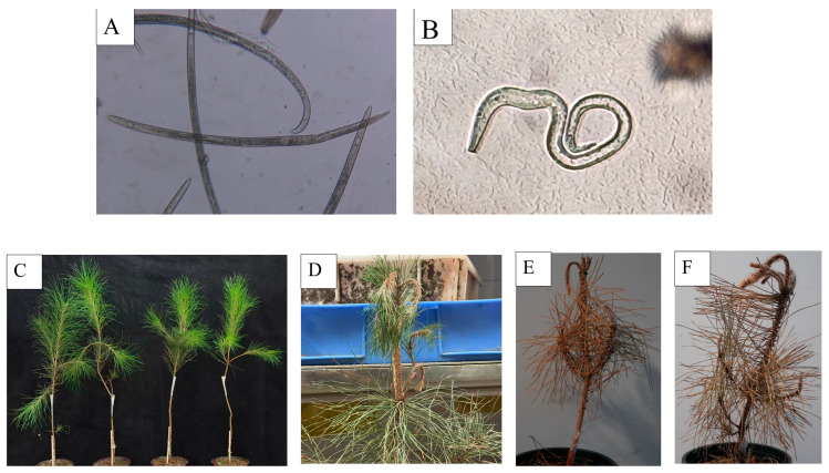
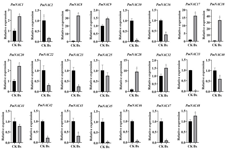
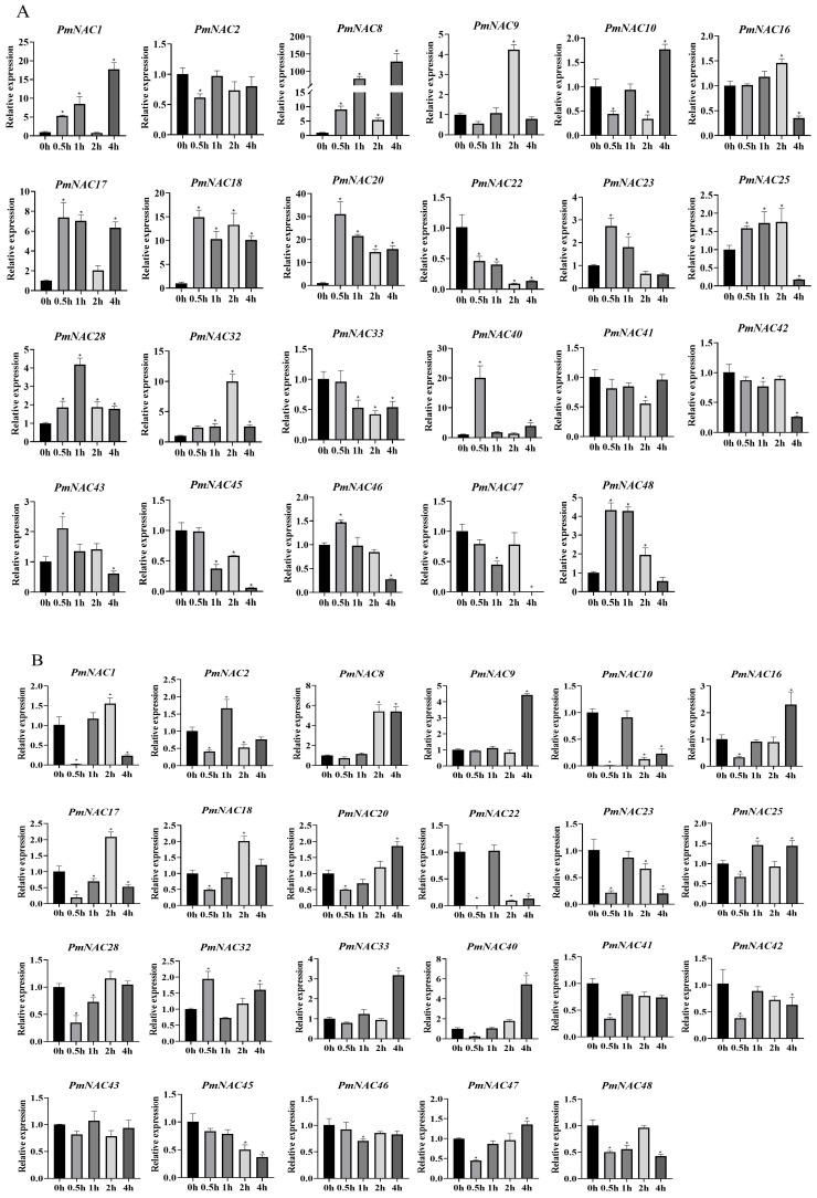
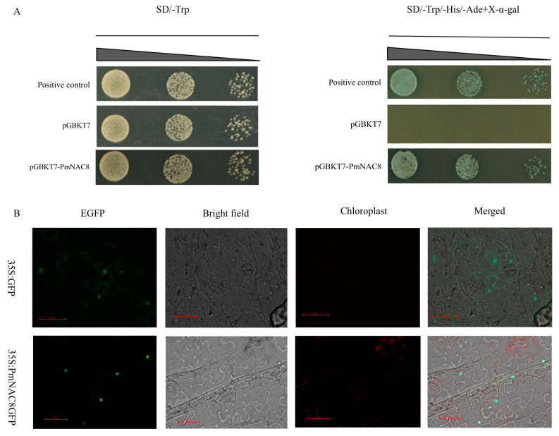
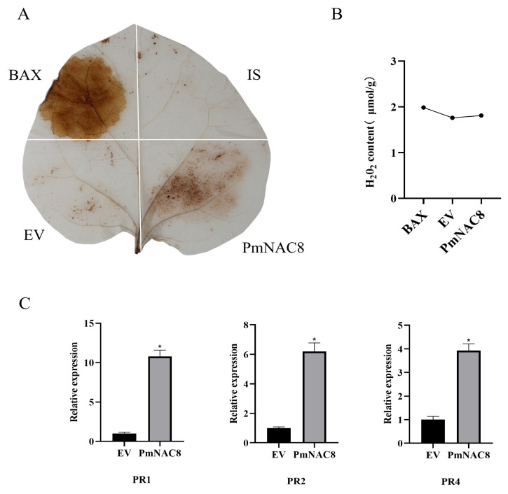
Pinus massoniana Lamb. is an economically important conifer native to China. However, it is highly susceptible to the pine wood nematode (Bursaphelenchus xylophilus, PWN), the causal agent of pine wilt disease (PWD), resulting in substantial ecological and economic losses. To elucidate potential molecular defense mechanisms, 50 NAC (NAM, ATAF1/2, and CUC2) transcription factors (PmNACs) were identified in the P. massoniana genome. Phylogenetic analysis divided these PmNACs into seven subfamilies, and motif analysis identified ten conserved motifs associated with stress responses. Twenty-three genes were selected for expression analysis in various tissues and under exogenous salicylic acid (SA), methyl jasmonate (MeJA), and PWN infection. Six genes (PmNAC1, PmNAC8, PmNAC9, PmNAC17, PmNAC18, and PmNAC20) were significantly up-regulated by both hormonal treatment and PWN infection, implying their involvement in JA/SA-mediated immune pathways. Functional characterization showed PmNAC8 is a nuclear-localized transcription factor with autoactivation activity. Furthermore, transient overexpression of PmNAC8 in Nicotiana benthamiana induced reactive oxygen species (ROS) accumulation and necrotic lesions. Collectively, these results elucidate NAC-mediated defense responses to PWN infection in P. massoniana and identify candidate genes for developing PWD-resistant pine varieties.

Keywords: Bursaphelenchus xylophilus; NAC family; hormones; pine wilt disease; subcellular localization; transcription factor.


Conflict of interest statement

The authors declare no conflicts of interest.

Figures

Figure 1
Figure 1
Multi-sequence alignment of conserved domains of Masson pine protein sequences. The red part indicates sequence similarity ≥ 75%, and the blue area indicates sequence similarity ≥ 50%. (AE) represent the five canonical structural subdomains of the NAC family. The blank area within the red box indicates that the gene is missing the corresponding fragment. Motif 3 constitutes subdomain A, Motif 5 constitutes subdomain B, Motif 2 and Motif 4 constitute subdomain C, Motif 1 and Motif 8 constitute subdomain D, and Motif 6 constitutes subdomain E.
Figure 2
Figure 2
Phylogenetic analysis of NAC genes from P. massoniana and A. thaliana. The unrooted phylogenetic tree was constructed using the Maximum Likelihood (ML) method with 1000 bootstrap replicates. NAC proteins from P. massoniana and A. thaliana are indicated by red and green dots, respectively. The seven major subfamilies of P. massoniana NACs are highlighted in different colored shades. Bootstrap values > 95% are shown on the corresponding branches.
Figure 3
Figure 3
Gene structure and motif analysis of PmNAC genes in P. massoniana. (A) The phylogenetic relationships among 50 NAC genes in P. massoniana were inferred using a Neighbor-Joining (NJ) tree. (B) Conserved protein motifs within the PmNAC genes are depicted as color-coded boxes, with each color representing a distinct motif.
Figure 4
Figure 4
Hierarchical clustering analysis of tissue-specific expression patterns of 23 PmNAC genes. Heatmap visualization of qRT-PCR normalized expression levels of PmNAC genes in root, stem, and leaf tissues. Heatmap of gene expression normalized to PmCYP (internal control) using the ΔCt method. Hierarchical clustering dendrogram based on Euclidean distance and average linkage. Color scale indicates relative expression levels (blue: low; red: high).
Figure 5
Figure 5
Morphological figures of B. xylophilus and changes in P. massoniana after inoculation with B. xylophilus. (A) Female. (B) Male. (C) The healthy P. massoniana plants were inoculated by B. xylophilus. (D) Two weeks after inoculation with B. xylophilus, the top of P. massoniana plants exhibited bending and wilting. (E,F) Four weeks after inoculation with B. xylophilus, the entire P. massoniana plant turned yellow and subsequently withered and died.
Figure 6
Figure 6
Differential expression of PmNAC genes in P. massoniana in response to B. xylophilus infection. The transcriptional response of 23 PmNAC genes to pine wood nematode (B. xylophilus) infection was investigated using RT-qPCR. Control samples (Control) were collected from mock-inoculated seedlings (treated with sterile water), while infected samples (BX) were collected from seedlings inoculated with nematodes. For each group, tissues from multiple whole seedlings were pooled. Relative gene expression was calculated using the 2−∆∆Ct method. Data are presented as the mean ± standard deviation (SD) of three biological replicates. Student’s t-test was used to determine statistical significance between the infected and control groups. Asterisks denote significant differences (*, p < 0.05).
Figure 7
Figure 7
Expression analysis of the PmNAC gene family in P. massoniana in response to phytohormone treatments. The relative expression levels of 23 PmNAC genes were analyzed in P. massoniana seedlings (or specify the tissue, e.g., needles) after 4 h of treatment. (A) Plants treated with 100 μM MeJA. (B) Plants treated with 5 mM SA. A mock treatment (e.g., with the solvent used to dissolve the hormones) served as the control group for both experiments. Gene expression was quantified using the 2−ΔΔCt method. Data are presented as the mean ± SD from three biological replicates. Asterisks indicate statistically significant differences compared to the respective mock-treated controls (* p < 0.05), as determined by Student’s t-test.
Figure 8
Figure 8
Analysis of PmNAC8 transcription factor characteristics. (A) The transcriptional activation activity of PmNAC8 was confirmed in yeast. Yeast cells transformed with a PmNAC8 construct grew on a selective triple-deficiency medium, demonstrating its function as a transcriptional activator. (B) Subcellular localization analysis in N. benthamiana epidermal cells showed that the PmNAC8-GFP fusion protein is localized to the nucleus, consistent with its function as a transcription factor. Scale bars = 50 µm.
Figure 9
Figure 9
Overexpression of PmNAC8 induces ROS accumulation and defense gene expression in N. benthamiana. (A) Visualization of hydrogen peroxide (H2O2) accumulation by 3,3′-diaminobenzidine (DAB) staining in N. benthamiana leaves at 48 h post-infiltration (hpi). Tissues were infiltrated with Agrobacterium carrying a PmNAC8 expression construct or an empty vector (EV) as a control. (B) Quantification of H2O2 content in the infiltrated leaf tissues. (C) Relative expression levels of pathogenesis-related (PR) genes in the infiltrated zones, as determined by RT-qPCR. Relative gene expression was calculated using the 2−∆∆Ct method. Data are presented as the mean ± standard deviation (SD) of three biological replicates. Student’s t-test was used to determine the statistical significance between the PmNAC8 injection group and the empty vector group. Asterisks indicate significant differences (*, p < 0.05).

Similar articles

References

    1. Chen J.-Q., Zhang L.-Z., Ma J., Li C.-J., Zang Y.-D., Sun H., Zhang D.-M. Hepatoprotective Diterpenes from the Nodes of Pinus Massoniana. J. Asian Nat. Prod. Res. 2025;27:995–1004. doi: 10.1080/10286020.2025.2506527. - DOI - PubMed
    1. Brugger D., Wilhelm B., Schusser B., Gisch N., Matthes J., Zhao J., Windisch W. Masson Pine Pollen (Pinus Massoniana) Activate HD11 Chicken Macrophages in Vitro. J. Ethnopharmacol. 2024;325:117870. doi: 10.1016/j.jep.2024.117870. - DOI - PubMed
    1. Tang F., Zhou Y., Feng J., Li J., Feng J., Lv Y., Mao Y., Wang Y., Deng P., Bai Y. Important Role of Pinus Massoniana Mixed Forests in Enhancing Soil Carbon Stocks in Degraded Forests in Southern China. CATENA. 2025;250:108792. doi: 10.1016/j.catena.2025.108792. - DOI
    1. Xu S., Huang W., Wang D., Zhang B., Sun H., Yan J., Ding J., Ma X. Risk Assessment of Carbon Stock Loss in Chinese Forests Due to Pine Wood Nematode Invasion. Forests. 2025;16:315. doi: 10.3390/f16020315. - DOI
    1. Modesto I., Mendes A., Carrasquinho I., Miguel C.M. Molecular Defense Response of Pine Trees (Pinus Spp.) to the Parasitic Nematode Bursaphelenchus Xylophilus. Cells. 2022;11:3208. doi: 10.3390/cells11203208. - DOI - PMC - PubMed

LinkOut - more resources