. 2025 Aug 7;14(15):2441.
doi: 10.3390/plants14152441.

A Novel Cysteine Protease from Phytolacca americana Cleaves Pokeweed Antiviral Protein Generating Bioactive Fragments

Affiliations

A Novel Cysteine Protease from Phytolacca americana Cleaves Pokeweed Antiviral Protein Generating Bioactive Fragments

Annabelle Audet et al. Plants (Basel). .

Abstract

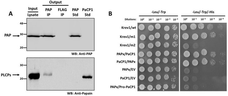
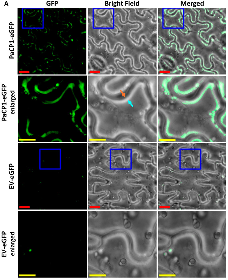
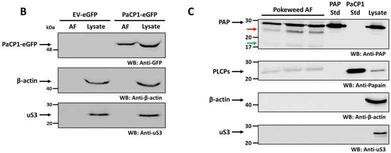
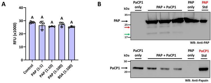
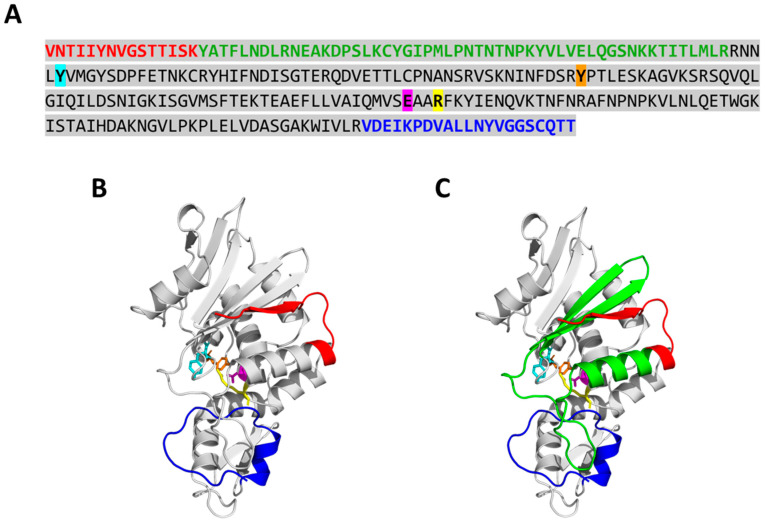
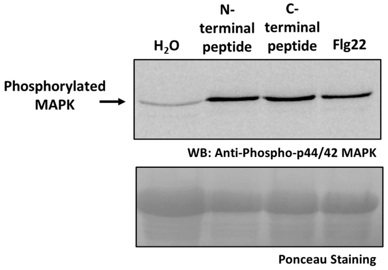
The apoplast is often the first point of contact between plant cells and invading pathogens, serving as an important site for defense signaling. Pokeweed antiviral protein (PAP), a ribosome-inactivating protein from Phytolacca americana (pokeweed), is localized to the apoplast and is hypothesized to accompany a pathogen to the cytosol, where it would inactivate host ribosomes to prevent pathogen spread. However, it is not known whether PAP interacts with other proteins in the apoplast. In this study, we identified Phytolacca americana cysteine protease 1 (PaCP1), an extracellular cysteine protease, as a novel PAP interactor. Sequence and structural analyses classified PaCP1 as a member of the C1A subfamily of papain-like cysteine proteases. Immunoprecipitation, mass spectrometry, and yeast two-hybrid analysis showed that PAP specifically binds the mature, active form of PaCP1. Curiously, PaCP1 cleaves PAP at its N- and C-termini, generating peptides that enhance MAPK phosphorylation in pokeweed leaves, indicating their potential role in stress signaling. PaCP1 processing of PAP to generate bioactive peptides diversifies the function of a ribosome-inactivating protein beyond its canonical inhibition of translation. Our findings present a novel extracellular role for PAP and advance our understanding of how protein interactions in the apoplast contribute to plant immune responses.

Keywords: RNA N-glycosylase; apoplast; cysteine protease; pokeweed antiviral protein; ribosome-inactivating protein.


Conflict of interest statement

The authors declare no conflicts of interest.

Figures

Figure 1
Figure 1
Protein sequence characterization of PaCP1. (A) The amino acid sequence for PaCP1 was aligned to the plant protein sequence with the highest homology, AtXCP1, using Blastp. Identical residues are shown in gray highlights and orange highlights indicate residues with similar chemical properties. Pink residues are required for recognition and cleavage of the signal peptide, and this cleavage site is indicated by the white arrow. Yellow residues are required for the recognition and cleavage of the propeptide inhibitor domain, and this cleavage site is indicated by the black arrow. The residues of the active site are colored in blue, red, and green and represent cysteine (C156), histidine (H291), and asparagine (N311), respectively. (B) Comparison schematic of the putative protein sequence organization for PaCP1 compared to the known protein organization of AtXCP1, obtained from UniProt (uniprot.org). The location of the signal peptide, propeptide inhibitor domain and peptidase C1A domain for PaCP1 were predicted using Interproscan, Signal-P 6.0, and Target-P 2.0. The active site residues for catalytic activity are cysteine (C; blue), histidine (H; red), and asparagine (N; green). Three disulfide bridges are shown in gray. aa: amino acids. (C) Structural prediction of the mature PaCP1 protein (peptidase C1A domain, residues 133–345) using the colab implementation of AlphaFold2. The three catalytic residues of the active site are colored as presented in (A,B).
Figure 2
Figure 2
Enzymatic activity of recombinant PaCP1. (A) Proteolytic activity of PaCP1 over a range of pH (4–7.5). RFU of the cleaved fluorescent substrate was measured after a 3 h incubation with the protease at 22 °C. Values are means ± standard deviation of n = 3 independent experiments. (B) Proteolytic activity of papain and PaCP1. Control samples represent an incubation with the fluorescent substrate and papain or PaCP1 only. The E-64 samples are supplemented with 100 μM E-64 specific inhibitor. Values are means ± standard deviation of n = 3 independent experiments. Different letters above the bars represent statistically significant differences. p values were calculated by two-way ANOVA (p < 0.001). (C) Enzyme kinetics of PaCP1 (0.1 µg) at varying FTC-Casein substrate concentrations (0.3 µM to 40.0 µM) after a 1 h incubation at 22 °C. Values are means ± standard deviation of n = 3 independent experiments.
Figure 3
Figure 3
Support for PAP-PaCP1 interaction. (A) Immunoblot of the immunoprecipitation assay using PAP to immunoprecipitate PLCPs in pokeweed. A FLAG immunoprecipitation serves as a negative control. The input lane represents total pokeweed protein from cell lysate. The PAP and FLAG output lanes represent the immunoprecipitated proteins. A PAP and PaCP1 standards are included. Proteins were detected by Western blot (WB) using anti-PAP and anti-papain antibodies. (B) Yeast-two hybrid assay testing the interaction between PAPx and PaCP1. Growth of yeast cells on -Leu/-Trp/-His media represent positive protein–protein interaction. The same cells were plated on -Leu/-Trp to confirm yeast transformation. Yeast transformed with empty vectors (EVs) served as negative activation control. Pro-PaCP1 is PaCP1 with its pro-inhibitory domain. Krev1/wt: strong positive interaction control. Krev1/m1: weak positive interaction control. Krev1/m2: negative interaction control.
Figure 4
Figure 4
PaCP1 and PAP localization to the extracellular space. (A) Fluorescence microscopy images showing PaCP1-eGFP localized to the extracellular space of N. benthamiana leaf epidermal cells, after salt-induced plasmolysis. EV-eGFP served as a negative control. Enlarged regions of interest (blue square) are shown below each original image. Red scale bars = 20 μM; yellow scale bars = 5 μM. Orange arrow indicates the plant cell wall, and the cyan arrow indicates the plasma membrane pulling away from the cell wall. (B) Western blot of apoplastic fluid (AF) and cell lysates from N. benthamiana leaves expressing PaCP1-eGFP or EV-eGFP, probed with an anti-GFP antibody. WB using anti-β-actin and anti-ribosomal protein uS3 antibodies served as controls for the absence of cytoplasmic protein contamination. (C) Western blot using anti-PAP and anti-papain antibodies to confirm presence of PAP and papain-like cysteine proteases in apoplastic fluid extracted from pokeweed leaves. Three samples of independent apoplastic fluid extractions from pokeweed are shown. The red and green arrows represent the lower molecular weight cleavage products of PAP. PAP and PaCP1 protein standards served as positive and/or negative controls. A sample of total pokeweed proteins was loaded in the far-right lane.
Figure 4
Figure 4
PaCP1 and PAP localization to the extracellular space. (A) Fluorescence microscopy images showing PaCP1-eGFP localized to the extracellular space of N. benthamiana leaf epidermal cells, after salt-induced plasmolysis. EV-eGFP served as a negative control. Enlarged regions of interest (blue square) are shown below each original image. Red scale bars = 20 μM; yellow scale bars = 5 μM. Orange arrow indicates the plant cell wall, and the cyan arrow indicates the plasma membrane pulling away from the cell wall. (B) Western blot of apoplastic fluid (AF) and cell lysates from N. benthamiana leaves expressing PaCP1-eGFP or EV-eGFP, probed with an anti-GFP antibody. WB using anti-β-actin and anti-ribosomal protein uS3 antibodies served as controls for the absence of cytoplasmic protein contamination. (C) Western blot using anti-PAP and anti-papain antibodies to confirm presence of PAP and papain-like cysteine proteases in apoplastic fluid extracted from pokeweed leaves. Three samples of independent apoplastic fluid extractions from pokeweed are shown. The red and green arrows represent the lower molecular weight cleavage products of PAP. PAP and PaCP1 protein standards served as positive and/or negative controls. A sample of total pokeweed proteins was loaded in the far-right lane.
Figure 5
Figure 5
PaCP1 cleaves PAP. (A) Enzymatic activity of recombinant PaCP1 in the presence of increasing amounts of PAP. Fluorescent substrate and PaCP1 were either alone (control) or were supplemented with increasing amounts of PAP (1:1, 1:10 and 1:100; PaCP1: PAP). BSA served as a negative control. RFU of the cleaved fluorescent substrate was measured after a 3 h incubation at 22 °C. Values are means ± standard deviation of n = 3 independent experiments. Different letters above the bars represent statistically significant differences. p values were calculated by one-way ANOVA (p > 0.05). (B) Western blot using anti-PAP showing PAP cleavage after 3 h incubation of PAP (10 μg) with 0.1 μg PaCP1 at 22 °C (three samples are shown). The red and green arrows represent the 24 kDa and 18 kDa cleavage products, respectively. Western blot using anti-papain antibody to confirm the presence of PaCP1 in the samples. PaCP1 alone and PAP alone were negative controls. A PAP and PaCP1 standard as a positive control in the far-right lanes.
Figure 6
Figure 6
Mass spectrometry analysis of PAP cleavage products. (A) Amino acid sequence of the 24 kDa and 18 kDa PAP cleavage products as identified by mass spectrometry. The amino acids colored in red (N-terminus) and blue (C-terminus) were missing from the 24 kDa PAP product. The amino acids in red and green (N-terminus) and blue (C-terminus) were missing from the 18 kDa PAP product. The amino acid sequence of mature full-length PAP is highlighted in gray. The amino acids of the PAP active site are colored as follows: tyrosine (Y72) in cyan, tyrosine (Y123) in orange, glutamic acid (E176) in pink, and arginine (R179) in yellow. (B,C) Structure of PAP obtained from the AlphaFold Protein Structure Database (AF-P10297-F1-v4) illustrating missing regions for the 24 (red and blue) and 18 kDa (red, green, and blue) product, respectively. The amino acids are colored the same as in (A).
Figure 7
Figure 7
Depurination activity of PAP cleavage products. qRT-PCR analysis of the levels of rRNA depurination of the 24 (24PAP) and 18 (18PAP) kDa PAP products. Purified N. benthamiana ribosomes (50 µg) were incubated with either 24PAP or 18PAP (5 µg), or full-length PAP for 30 min at 30 °C followed by rRNA isolation. qRT-PCR measured the levels of reference and target 25S rRNA products relative to samples treated with buffer alone. Different letters above the bars represent statistically significant differences. Values are means ± standard deviation of n = 3 independent experiments. p values were calculated using one-way ANOVA (p <0.01).
Figure 8
Figure 8
Increase in MAPK phosphorylation by N- and C-terminal peptides of PAP. Immunoblot using anti-phospho p44/42 MAPK polyclonal antibody showing MAPK phosphorylation in protein lysates (35 μg) from pokeweed leaves infiltrated with either N-terminal or C-terminal peptide from the 24 kDa PAP cleavage product. Leaves infiltrated with water or Flg22 served as negative and positive controls, respectively. The blot was stained with Ponceau to illustrate loading amounts.

Similar articles

References

    1. Sattelmacher B. The apoplast and its significance for plant mineral nutrition. New Phytol. 2001;149:167–192. doi: 10.1046/j.1469-8137.2001.00034.x. - DOI - PubMed
    1. Farvardin A., González-Hernández A.I., Llorens E., García-Agustín P., Scalschi L., Vicedo B. The Apoplast: A Key Player in Plant Survival. Antioxidants. 2020;9:604. doi: 10.3390/antiox9070604. - DOI - PMC - PubMed
    1. Darino M., Kanyuka K., Hammond-Kosack K.E. Apoplastic and vascular defences. Essays Biochem. 2022;66:595–605. doi: 10.1042/ebc20220159. - DOI - PubMed
    1. Qi J., Wang J., Gong Z., Zhou J.-M. Apoplastic ROS signaling in plant immunity. Curr. Opin. Plant Biol. 2017;38:92–100. doi: 10.1016/j.pbi.2017.04.022. - DOI - PubMed
    1. Miedes E., Vanholme R., Boerjan W., Molina A. The role of the secondary cell wall in plant resistance to pathogens. Front. Plant Sci. 2014;5:358. doi: 10.3389/fpls.2014.00358. - DOI - PMC - PubMed

LinkOut - more resources