. 2025 Jul 28;13(15):1838.
doi: 10.3390/healthcare13151838.

Continuous Radiofrequency for Morton's Neuroma: Is There Complete Ablation? A Preliminary Report

Affiliations

Continuous Radiofrequency for Morton's Neuroma: Is There Complete Ablation? A Preliminary Report

Gabriel Camuñas-Nieves et al. Healthcare (Basel). .

Abstract

Background and Objectives: Morton's neuroma is a painful foot condition that can be treated with continuous radiofrequency. However, its efficacy is not always optimal, with failure rates of 15-20%. It has been suggested that these failures may be due to incomplete nerve ablation, allowing for nerve regeneration and persistent pain. So, the aim of this study was to assess the histological effects of continuous radiofrequency on the nerves affected by Morton's neuroma. Materials and Methods: The effect of continuous radiofrequency was evaluated in two patients with Morton's neuroma, which required open surgery excision. In both cases, radiofrequency with a standard protocol was applied ex vivo, following the surgical excision of the neuroma. A TLG10 RF generator (90 °C, 90 s) with a monopolar needle with a 0.5 cm active tip was used. Subsequently, the samples were histologically analyzed to determine the degree of nerve ablation. Results: Histological analysis showed homogeneous focal necrosis in both cases, with lesion depths of 2.4 mm and 3.18 mm. However, areas of intact nerve tissue were identified at the periphery of the neuroma, suggesting incomplete ablation. Conclusions: The findings indicate that continuous radiofrequency does not guarantee total nerve ablation, which could explain recurrence in some cases. Intraoperative neurophysiological monitoring could be key to optimizing the procedure, ensuring complete interruption of nerve conduction and improving treatment efficacy.

Keywords: Morton’s neuroma; histological analysis; nerve ablation; neuroma; radiofrequency.


Conflict of interest statement

The authors declare no conflicts of interest.

Figures

Figure 1
Figure 1
Ultrasound scans of a Morton’s neuroma in the third intermetatarsal space. Top: Case 1. Longitudinal (left) and transverse (right) ultrasound views showing a neuroma measuring 1.18 × 0.67 cm. Bottom: Case 2. Longitudinal (left) and transverse (right) ultrasound views showing a neuroma measuring 0.74 × 0.38 cm. In both cases, a well-defined hypoechoic structure is visualized between the metatarsal heads.
Figure 2
Figure 2
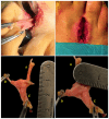
Intraoperative and anatomical images corresponding to a Morton’s neuroma in the third intermetatarsal space following surgical excision (Case 1). Top: Surgical exposure of the interdigital nerve via a dorsal approach. Bottom: Macroscopic view of the neuroma with three identifiable nerve branches. Three main nerve branches are identified: A: third interdigital branch. B: medial plantar digital. C: third intermetatarsal common nerve.
Figure 3
Figure 3
Ex vivo continuous radiofrequency procedure on a Morton’s neuroma. (A) Macroscopic image of the specimen after surgical excision, with longitudinal measurement of the neuroma using a millimeter scale. (B) Radiofrequency application using an active needle positioned directly on the neuroma. The metallic background corresponds to the return plate of the generator, which was required to complete the electrical circuit and enable thermal transmission during the ablation process.
Figure 4
Figure 4
Histological sections of a Morton’s neuroma following radiofrequency application, stained with hematoxylin and eosin. The darker purple regions represent areas where radiofrequency caused the most significant thermal damage, resulting in substantial ablation of nervous tissue. (A) Panoramic section from Case 1 showing partial thermal necrosis up to 2.4 mm in depth. (B) Panoramic section from Case 2 demonstrating more extensive necrosis reaching 3.18 mm, with a reduced amount of viable neural tissue. (C) Magnified image from Case 1 showing disorganized nerve fascicles, perineural fibrosis, and tissue degeneration consistent with thermally induced damage.

References

    1. Mann R.A., Reynolds J.C. Interdigital neuroma—A critical clinical analysis. Foot Ankle. 1983;3:238–243. doi: 10.1177/107110078300300411. - DOI - PubMed
    1. Adams W.R., 2nd Morton’s neuroma. Clin. Podiatr. Med. Surg. 2010;27:535–545. doi: 10.1016/j.cpm.2010.06.004. - DOI - PubMed
    1. Wu K.K. Morton’s interdigital neuroma: A clinical review of its etiology, treatment, and results. J. Foot Ankle Surg. 1996;35:112–119. doi: 10.1016/S1067-2516(96)80027-5. discussion 187–188. - DOI - PubMed
    1. Valisena S., Petri G.J., Ferrero A. Treatment of Morton’s neuroma: A systematic review. Foot Ankle Surg. 2018;24:271–281. doi: 10.1016/j.fas.2017.03.010. - DOI - PubMed
    1. Ruiz Santiago F., Prados Olleta N., Tomas Munoz P., Guzman Alvarez L., Martinez Martinez A. Short term comparison between blind and ultrasound guided injection in morton neuroma. Eur. Radiol. 2019;29:620–627. doi: 10.1007/s00330-018-5670-1. - DOI - PubMed

LinkOut - more resources