. 2025 Jul 24;26(15):7122.
doi: 10.3390/ijms26157122.

Electromagnetic Transduction Therapy (EMTT) Enhances Tenocyte Regenerative Potential: Evidence for Senolytic-like Effects and Matrix Remodeling

Affiliations

Electromagnetic Transduction Therapy (EMTT) Enhances Tenocyte Regenerative Potential: Evidence for Senolytic-like Effects and Matrix Remodeling

Matteo Mancini et al. Int J Mol Sci. .

Abstract

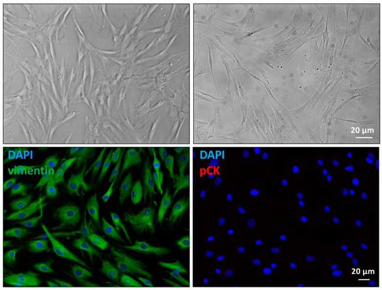
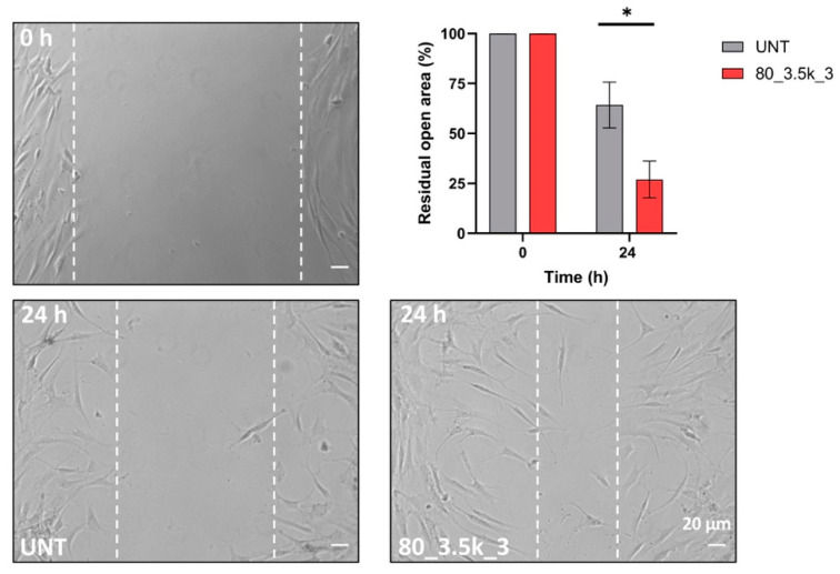
Tendinopathies are a significant challenge in musculoskeletal medicine, with current treatments showing variable efficacy. Electromagnetic transduction therapy (EMTT) has emerged as a promising therapeutic approach, but its biological effects on tendon cells remain largely unexplored. Here, we investigated the effects of EMTT on primary cultured human tenocytes' behavior and functions in vitro, focusing on cellular responses, senescence-related pathways, and molecular mechanisms. Primary cultures of human tenocytes were established from semitendinosus tendon biopsies of patients undergoing anterior cruciate ligament (ACL) reconstruction (n = 6, males aged 17-37 years). Cells were exposed to EMTT at different intensities (40 and 80 mT) and impulse numbers (1000-10,500). Cell viability (MTT assay), proliferation (Ki67), senescence markers (CDKN2a/INK4a), migration (scratch test), cytoskeleton organization (immunofluorescence), and gene expression (RT-PCR) were analyzed. A 40 mT exposure elicited minimal effects, whereas 80 mT treatments induced significant cellular responses. Repeated 80 mT exposure demonstrated a dual effect: despite a moderate decrease in overall cell vitality, increased Ki67 expression (+7%, p ≤ 0.05) and significant downregulation of senescence marker CDKN2a/INK4a were observed, suggesting potential senolytic-like activity. EMTT significantly enhanced cell migration (p < 0.001) and triggered cytoskeletal remodeling, with amplified stress fiber formation and paxillin redistribution. Molecular analysis revealed upregulation of tenogenic markers (Scleraxis, Tenomodulin) and enhanced Collagen I and III expressions, particularly with treatments at 80 mT, indicating improved matrix remodeling capacity. EMTT significantly promotes tenocyte proliferation, migration, and matrix production, while simultaneously exhibiting senolytic-like effects through downregulation of senescence-associated markers. These results support EMTT as a promising therapeutic approach for the management of tendinopathies through multiple regenerative mechanisms, though further studies are needed to validate these effects in vivo.

Keywords: EMTT; cell migration; extracellular matrix remodeling; human tenocytes; mechanotransduction; senescence; senolytic; tendinopathy; tendon regeneration.


Conflict of interest statement

The authors declare that they have no conflicts of interest.

Figures

Figure 1
Figure 1
Images of the primary culture of human tenocytes. Top panels, differential interference contrast (left) and phase contrast microscopy (right). Bottom panels, cells that were stained with 4′,6-diamidino-2-phenylindole (DAPI) and vimentin (left), or human pan-cytokeratin (pCK) (right).
Figure 2
Figure 2
Experimental design of the dose–response study (T1 = 48 h; T2 = 54 h; T3 = 55 h). (a) Two field intensities approach. (b) Higher field intensities with variable number of pulses approach. Created with https://BioRender.com on 27 April 2025.
Figure 3
Figure 3
Cell viability and proliferation evaluation in EMTT-treated cells. (a) Cells treated as described above were evaluated for vitality by colorimetric MTT assay. (b) Cells treated as above were stained for immunofluorescence, and nuclei were visualized by DAPI staining. Bar: The proliferation rate, assessed by immunofluorescence with an anti-Ki67 polyclonal antibody, reveals positive cycling cells comparable in all the experimental conditions; 20 µm. The results are expressed as mean fold increase ± standard deviation (SD), * p < 0.05, ** p < 0.01, *** p < 0.001, **** p < 0.0001. n.s. means not significant.
Figure 4
Figure 4
Modulation of mRNA expression levels of differentiation markers after EMTT exposure. (ah) Bar graphs showing gene expression levels of the different markers: scleraxis, SCX (a); tenomodulin, TNMD (b); tenascin-C, TNC (c); type I collagen, COL1A1 (d); type III collagen, COL3A1 (e); alpha smooth muscle actin (α-SMA), ACTA2 (f); cyclin-dependent kinase inhibitor 2A (p16), CDKN2A (g); matrix metalloproteinase-9, MMP9 (h) in cells treated as above compared to untreated cells (UNT). Results are expressed as mean value ± SD. ns, not statistically significant; * p < 0.05; ** p < 0.01; *** p < 0.001, **** p < 0.0001.
Figure 5
Figure 5
Effects of EMTT exposure on tenocytes’ migration abilities. Representative images of scratch assay (n = 3) showing the effect of EMTT on cell migration. Bar: 20 µm. The percentage of residual open area after 24 h of treatment with EMTT at 80_3.5k_3, compared to that of untreated cells (UNT), was measured using the Axiovision software as reported in the Materials and Methods section. Results are expressed as mean value ± SD, * p < 0.05.
Figure 6
Figure 6
Immunofluorescence analysis of the effects of acute EMTT exposure on actin cytoskeleton architecture and paxillin expression in tenocytes. Representative IF acquisitions (n = 3) showing actin cytoskeleton (top panels) and paxillin expression (bottom panels) of cells treated with EMTT at 80_3.5k_3 or untreated (UNT). F-Actin is stained in red with TRITC–Phalloidin, paxillin is stained in green with FITC-conjugated antibody, and nuclei are stained in blue with DAPI. Bar: 10 µm. Quantitative immunofluorescence analysis of TRITC–Phalloidin (top graph) and FITC–paxillin (bottom graph) staining is expressed as the mean ± SD relative fluorescence unit normalized to untreated cells (* p < 0.05; ** p < 0.01).

References

    1. Sharma P., Maffulli N. Tendinopathy and tendon injury: The future. Disabil. Rehabil. 2008;30:1733–1745. doi: 10.1080/09638280701788274. - DOI - PubMed
    1. Hopkins C., Fu S.C., Chua E., Hu X., Rolf C., Mattila V.M., Qin L., Yung P.S.-H., Chan K.-M. Critical review on the socioeconomic impact of tendinopathy. Asia Pac. J. Sports Med. Arthrosc. Rehabil. Technol. 2016;4:9–20. doi: 10.1016/j.asmart.2016.01.002. - DOI - PMC - PubMed
    1. Maffulli N., Khan K.M., Puddu G. Overuse tendon conditions: Time to change a confusing terminology. Arthroscopy. 1998;14:840–843. doi: 10.1016/S0749-8063(98)70021-0. - DOI - PubMed
    1. Scott A., Squier K., Alfredson H., Bahr R., Cook J.L., Coombes B., de Vos R.-J., Fu S.N., Grimaldi A., Lewis J.S., et al. ICON 2019: International Scientific Tendinopathy Symposium Consensus: Clinical Terminology. Br. J. Sports Med. 2020;54:260–262. doi: 10.1136/bjsports-2019-100885. - DOI - PubMed
    1. Chisari E., Rehak L., Khan W.S., Maffulli N. Tendon healing in presence of chronic low-level inflammation: A systematic review. Br. Med. Bull. 2019;132:97–116. doi: 10.1093/bmb/ldz035. - DOI - PubMed

LinkOut - more resources