. 2025 Jul 28;26(15):7288.
doi: 10.3390/ijms26157288.

PKC- ι Regulates an Oncogenic Positive Feedback Loop Between the MAPK/JNK Signaling Pathway, c-Jun/AP-1 and TNF- α in Breast Cancer

Affiliations

PKC- ι Regulates an Oncogenic Positive Feedback Loop Between the MAPK/JNK Signaling Pathway, c-Jun/AP-1 and TNF- α in Breast Cancer

Nuzhat Nowshin Oishee et al. Int J Mol Sci. .

Abstract

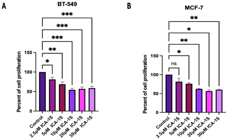
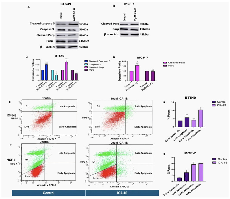
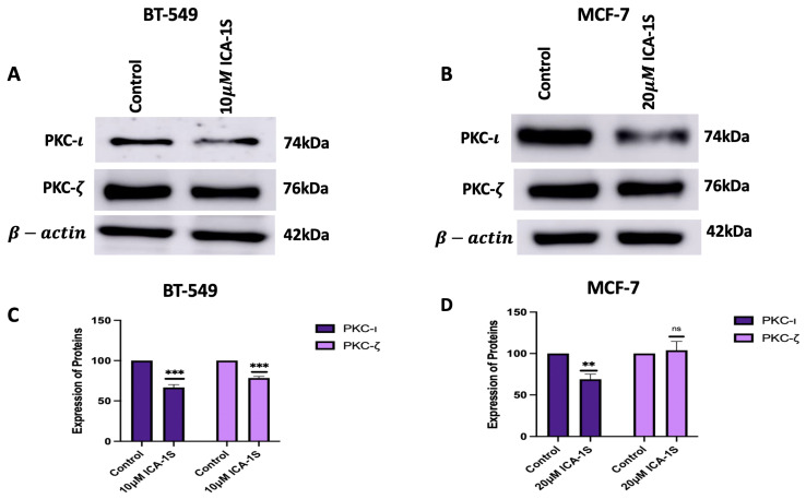
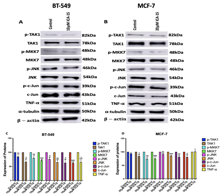
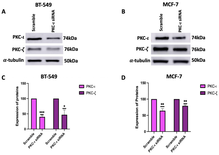
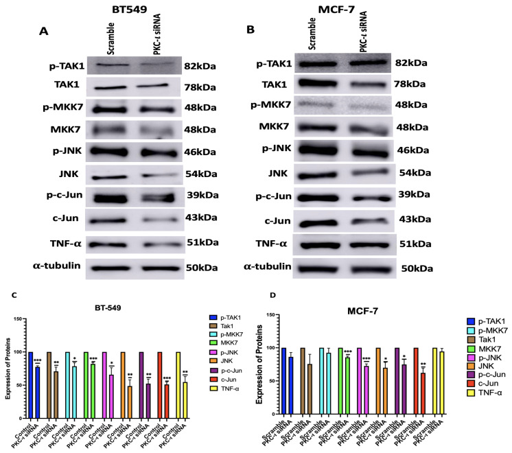
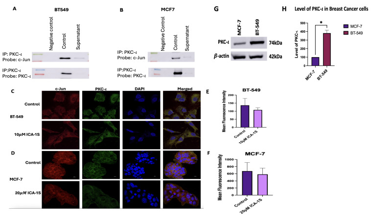
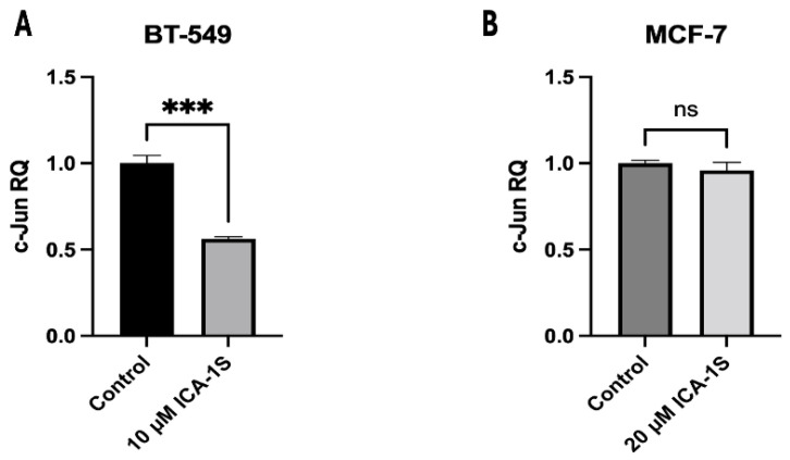
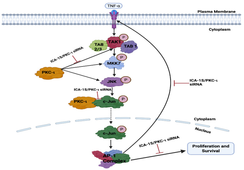
Breast cancer is the second most common cancer in the United States and consists of 30% of all new female cancer each year. PKC iota (PKC-ι) is a bonafide human oncogene and is overexpressed in many types of cancer, including breast cancer. This study explores the role of PKC-ι in regulating the transcription factor Jun proto-oncogene (c-Jun), pro-inflammatory cytokine Tumor Necrosis Factor-alpha (TNF-α), and the Mitogen-Activated Protein Kinase/Jun N-terminal kinase (MAPK/JNK) pathway, which also exhibits an oncogenic role in breast cancer. ICA-1S, a PKC-ι specific inhibitor, was used to inhibit PKC-ι to observe the subsequent effect on the levels of c-Jun, TNF-α, and the MAPK/JNK signaling pathway. To obtain the results, cell proliferation assay, Western blotting, co-immunoprecipitation, small interfering RNA (siRNA), immunofluorescence, flow cytometry, cycloheximide (CHX) chase assay, and reverse transcription quantitative PCR (RT-qPCR) techniques were implemented. ICA-1S significantly inhibited cell proliferation and induced apoptosis in both breast cancer cell lines. Treatment with ICA-1S and siRNA also reduced the expression levels of the MAPK/JNK pathway protein, c-Jun, and TNF-α in both cell lines. PKC-ι was also found to be strongly associated with c-Jun, via which it regulated the MAPK/JNK pathway. Additionally, ICA-1S was found to promote the degradation of c-Jun and decrease the mRNA levels of c-Jun. We concluded that PKC-ι plays a crucial role in regulating breast cancer, and the inhibition of PKC-ι by ICA-1S reduces breast cancer cell proliferation and induces apoptosis. Therefore, targeting PKC-ι as a potential therapeutic target in breast cancer could be a significant approach in breast cancer research.

Keywords: MAPK/JNK pathway; TNF-α; apoptosis; breast cancer; c-Jun.


Conflict of interest statement

The authors declare no conflicts of interest. The funders had no role in the design of this study; in the collection, analysis, or interpretation of data; in the writing of this manuscript; or in the decision to publish the results.

Figures

Figure 1
Figure 1
Dose response curve of ICA-1S in (A) BT-549 and (B) MCF-7 cell line. The results for the cell proliferation assay indicates ICA-1S has an inhibitory effect on the cell proliferation of breast cancer cell lines. The graph represents the mean ± Standard Error of Mean (SEM) of 3 independent experiments of each cell line. *** p < 0.001; ** p < 0.005, * p < 0.05, ns: not significant.
Figure 2
Figure 2
Treatment with ICA-1S induces apoptosis for both BT-549 and MCF-7 cells. (AD) Representative Western blot images and the corresponding graph indicate that treatment with ICA-1S increases the levels of cleaved Caspase 3 and cleaved PARP, which are hallmark events in apoptosis. Expression levels were normalized to the corresponding loading controls such as β-actin and α-tubulin. (EH) Flow cytometry analysis using Annexin V-APC/PI staining demonstrates an increase in both early and late apoptotic cell populations with ICA-1S treatment when compared to the control group (untreated group) as observed in the individual cell dot plots for both BT-549 and MCF-7 cells. The graph represents the mean ± Standard Error of Mean (SEM) of 3 independent experiments of each cell line. *** p < 0.001; ** p < 0.005, * p < 0.05. ns: not significant.
Figure 3
Figure 3
Representative (A,C) represents the Western blot images and its corresponding graphs for BT-549 cells and (B,D) represents the western blot images and its corresponding graph for MCF-7 cells. The results demonstrate the treatment with ICA-1S decreases the levels of PKC-ι in both BT-549 and MCF-7 cells, while the level of PKC-ζ was decreased in BT-549 cells but increased in MCF-7 cells. Expression levels were normalized to the corresponding loading controls such as β-actin and α-tubulin. The graph represents the Mean ± Standard Error of Mean (SEM) of 3 independent experiments of each cell line. *** p < 0.001; ** p < 0.005, ns: not significant.
Figure 4
Figure 4
(A,B) Western blot images and (C,D) corresponding graphical representation demonstrating the effect of ICA-1S on the phosphorylation levels and the total protein level of the MAPK/JNK pathway proteins for BT-549 and MCF-7 cells. Expression levels were normalized to the corresponding loading controls such as β-actin and α-tubulin. The graph represents the Mean ± Standard Error of Mean (SEM) of 3 independent experiments of each cell line. *** p < 0.001; ** p < 0.005, * p < 0.05.
Figure 5
Figure 5
(A,B) Western blot images and (C,D) corresponding graphical representation demonstrating effect of PKC-ι knockdown on aPKCs in BT-549 and MCF-7 cells. Treatment with PKC-ι siRNA lowered the levels of PKC-ι in both cell lines, as well as lowered the levels of PKC-ζ in both cell lines. Expression levels were normalized to the corresponding loading controls such as α-tubulin. The graph represents the Mean ± Standard Error of Mean (SEM) of 3 independent experiments of each cell line. *** p < 0.001; ** p < 0.005, * p < 0.05.
Figure 6
Figure 6
(A,B) Western blot images and (C,D) corresponding graphical representation demonstrating the effect PKC-ι knockdown on the expression of MAPK-JNK pathway proteins and their phosphorylated form in BT-549 and MCF-7 cells. Expression levels were normalized to the corresponding loading controls such as β-actin and α-tubulin. Graphs represents the Mean ± Standard Error of Mean (SEM) of 3 independent experiments of each cell line. *** p < 0.001; ** p < 0.005, * p < 0.05.
Figure 7
Figure 7
(A,B) Co-immunoprecipitation of c-Jun with PKC-ι in BT-549 and MCF-7 cell line. Representative Western blot images indicate that there is a direct association between PKC-ι and c-Jun for both the cell lines. (CF) Immunofluorescence staining and visualization at 100X magnification of c-Jun protein with 4’,6-diamidino-2-phenylindole (DAPI) staining in BT-549 and MCF-7 cell lines. Treatment with ICA-1S also reduces the mean fluorescence intensity of c-Jun when compared to the control. (G,H) Level of PKC-ι in MCF-7 and BT-549 cells. MCF-7 cells demonstrate lower levels of PKC-ι, compared to BT-549 cells. The graph represents the Mean ± Standard Error of Mean (SEM) of 3 independent experiments of each cell line. * p < 0.05.
Figure 8
Figure 8
(A,B) Western blot images and (C,D) corresponding graphical representation demonstrating the expression of c-Jun protein with and without CHX treatment for BT-549 and MCF-7 cells. An enhanced decrease in the levels of c-Jun is observed after CHX treatment in the ICA-1S treatment group. Expression levels are normalized to the corresponding loading control such as α-tubulin. Graphs represents the Mean ± Standard Error of Mean (SEM) of 3 independent experiments of each cell line. *** p < 0.001; ** p < 0.005, * p < 0.05.
Figure 9
Figure 9
Expression of c-Jun mRNA in (A) BT-549 and (B) MCF-7 cell line. The graph representing BT-549 cells demonstrate the relative quantification value (RQ) of the target gene (c-Jun) is 0.5, which indicates a twofold downregulation in comparison to the control group, following normalization with the reference gene, GAPDH. However, no significant downregulation was observed for MCF-7 cells. Graphs represent the Mean ± Standard Error of Mean (SEM) of 3 independent experiments of each cell line. *** p < 0.001, ns: not significant.
Figure 10
Figure 10
Schematic representation of the possible pathway via which PKC-ι regulates MAPK/JNK pathway proteins in breast cancer. The inhibition of PKC-ι leads to c-Jun degradation or reduced expression, which decreases the AP-1 activity. Decrease in AP-1 activity reduces the levels of TNF-α, TAK-1, MKK7 and JNK. Decrease in the levels of JNK activity further decreases c-Jun expression, thereby reinforcing the oncogenic positive feedback loop. All these proteins play an oncogenic role in breast cancer proliferation and survival; therefore, reducing the levels of these proteins will lead to decreased proliferation and survival.

Similar articles

References

    1. World Health Organization Breast Cancer. 2024. [(accessed on 30 May 2025)]. Available online: https://www.who.int/news-room/fact-sheets/detail/breast-cancer.
    1. Garg R., Benedetti L.G., Abera M.B., Wang H., Abba M., Kazanietz M.G. Protein kinase C and cancer: What we know and what we do not. Oncogene. 2014;33:5225–5237. doi: 10.1038/onc.2013.524. - DOI - PMC - PubMed
    1. Marengo B., De Ciucis C.G., Ricciarelli R., Pronzato M.A., Marinari U.M., Domenicotti C. Protein kinase C: An attractive target for cancer therapy. Cancers. 2011;3:531–567. doi: 10.3390/cancers3010531. - DOI - PMC - PubMed
    1. Webb B.L., Hirst S.J., Giembycz M.A. Protein kinase C isoenzymes: A review of their structure, regulation and role in regulating airways smooth muscle tone and mitogenesis. Br. J. Pharmacol. 2000;130:1433–1452. doi: 10.1038/sj.bjp.0703452. - DOI - PMC - PubMed
    1. Kojima Y., Akimoto K., Nagashima Y., Ishiguro H., Shirai S., Chishima T., Ichikawa Y., Ishikawa T., Sasaki T., Kubota Y., et al. The overexpression and altered localization of the atypical protein kinase C lambda/iota in breast cancer correlates with the pathologic type of these tumors. Hum. Pathol. 2008;39:824–831. doi: 10.1016/j.humpath.2007.11.001. - DOI - PubMed

LinkOut - more resources