. 2025 Jul 28;26(15):7297.
doi: 10.3390/ijms26157297.

Membrane Progesterone Receptor Beta Regulates the Decidualization of Endometrial Stromal Cells in Women with Endometriosis

Affiliations

Membrane Progesterone Receptor Beta Regulates the Decidualization of Endometrial Stromal Cells in Women with Endometriosis

Dora Maria Velázquez-Hernández et al. Int J Mol Sci. .

Abstract

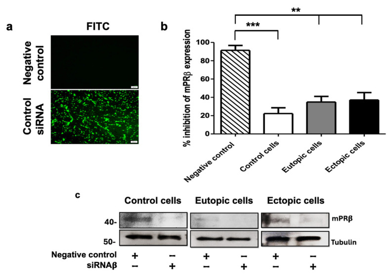
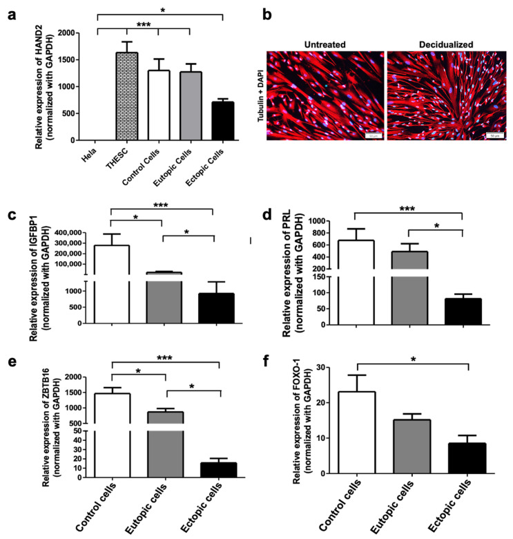
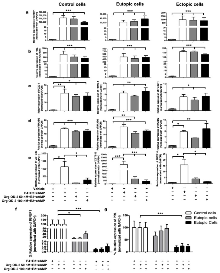
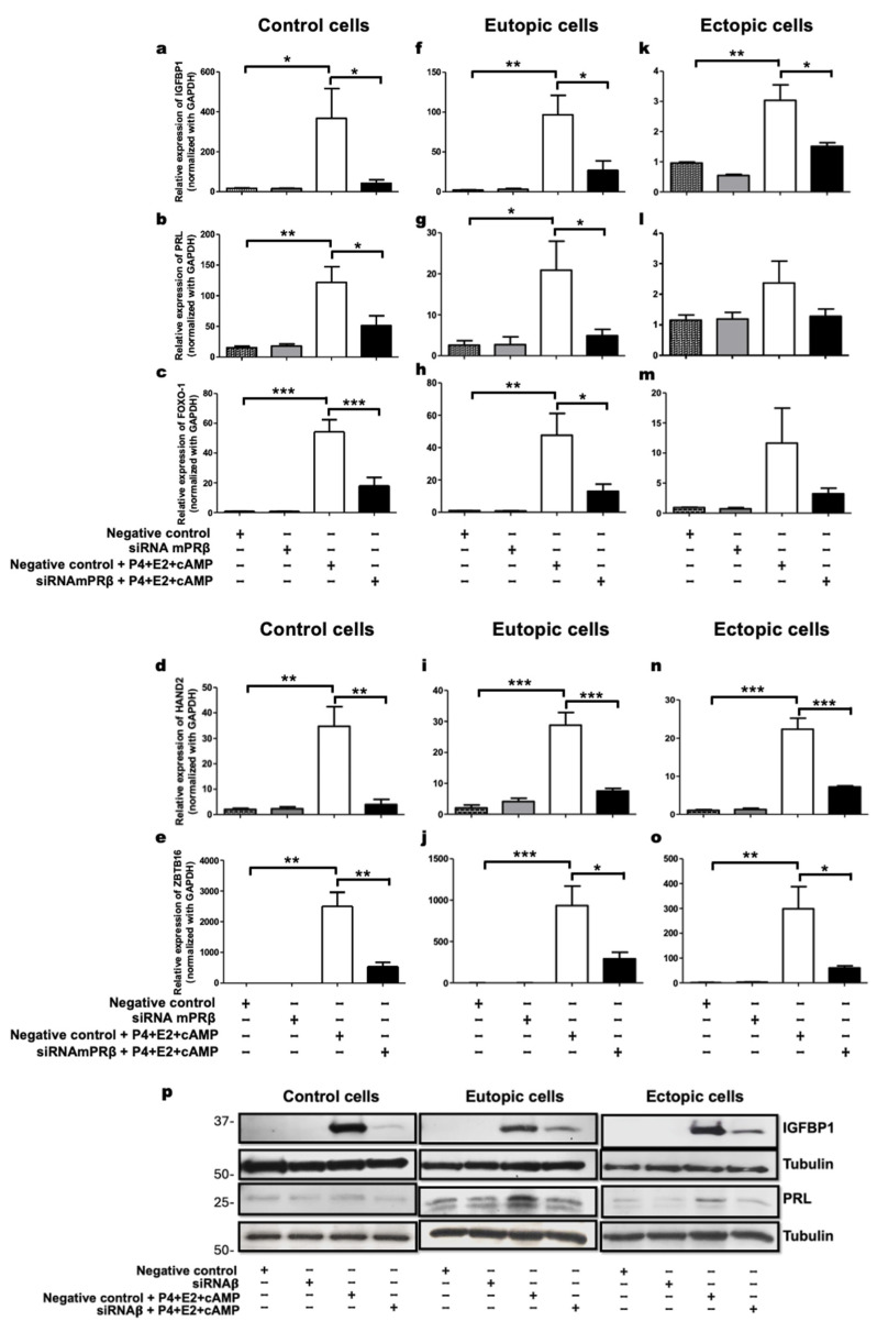
Endometriosis is a disorder characterized by the presence of endometrial tissue outside the uterus, leading to dyspareunia, chronic pelvic pain, dysuria, and infertility. The latter has been related to implantation failure associated with alterations in decidualization, a process regulated by sex hormones such as progesterone. Membrane progesterone receptor β (mPRβ) exhibits a lower expression in endometriotic tissues than in normal endometrial ones. However, the role of mPRβ in decidualization is unknown. This work aimed to investigate whether mPRβ plays a role in the decidualization of endometrial stromal cells (ESCs) derived from women with and without endometriosis. The mPR agonist OrgOD-2 induced the gene expression of key decidualization markers (insulin-like growth factor binding protein 1, prolactin, transcription factor heart and neural crest derivatives-expressed transcript 2, and fork-head transcription factor) in healthy ESCs, eutopic (uterine cavity), and ectopic (outside of the uterine cavity) ESCs from women with endometriosis. Notably, the expression of the decidualization markers was lower in endometriotic cells than in healthy endometrial ones. An siRNA mediated knockdown of mPRβ reduced the expression of decidualization-associated genes in ESCs treated with a decidualization stimuli, regardless of whether cells were derived from healthy women or those with endometriosis. Our data suggest that progesterone, through mPRβ activation, regulates the decidualization process in endometrial stromal cells from women with and without endometriosis.

Keywords: decidualization; endometrial stromal cells; endometriosis; membrane progesterone receptors; progesterone.


Conflict of interest statement

The authors declare no conflicts of interest.

Figures

Figure A1
Figure A1
Transfection of control and mPRβ siRNA into human endometrial stromal cells. (a) Representative image of control cells transfected with control siRNA (FITC conjugate) for 7 h, scale bar: 100 μm. (b) Control, eutopic, and ectopic cells were transfected with mPRβ siRNA or negative control for 48 h. The percentage of transfection was evaluated by the expression of mPRβ using RT-qPCR. The data in the figures represent mean ± SEM obtained from 10 independent experiments. Statistical analysis was performed using a one-way analysis of variance (ANOVA) followed by the Tukey post hoc test. Statistical significance was considered as follows: ** p ≤ 0.01 and *** p ≤ 0.001. (c) Representative image of protein levels of mPRβ in control, eutopic, and ectopic transfected cells with mPRβ siRNA or negative control for 48 h.
Figure 1
Figure 1
Decidualization of endometrial stromal cells from women with endometriosis. (a) Expression of HAND2 in various cell types: Hela cells of epithelial origin, THESC cell line derived from the endometrium of healthy women, control cells (women without endometriosis), eutopic cells (obtained from endometriotic lesions in the uterine cavity), and ectopic cells (obtained from endometriotic lesions outside the uterine cavity). (b) Immunofluorescence: representative image of the decidualization of control cells treated with the decidualization cocktail (E2 + cAMP + P4) scale bar 50 μm. (cf) Expression of decidualization markers IGFBP1, PRL, ZBTB16, and FOXO1 in control, eutopic, and ectopic cells treated with the decidualization cocktail for 48 h. Data in the figures represent mean + SEM obtained from 10 independent experiments. (a) Statistical analysis was performed using one-way analysis of variance (ANOVA) followed by the Tukey post hoc test. (cf) Statistical analysis was performed using Kruskal–Wallis followed by Dunns’s Multiple Comparison test. Statistical significance was considered as follows: * p ≤ 0.05 and *** p ≤ 0.001.
Figure 2
Figure 2
mPRs participate in the decidualization of endometrial stromal cells from women with and without endometriosis. (a) Expression of IGFBP1 and (b), PRL (c), FOXO1, (d) HAND2, and (e) ZBTB16 in control, eutopic, and ectopic cells treated with vehicle, 50 nM and 100 nM of the selective mPR agonist Org OD2 + 10 nM E2 + 0.5 mM cAMP. The graphs (f,g) depict the comparison in percentage of expression levels of decidualization markers IGFBP1 and PRL among control cells (women without endometriosis), eutopic, and ectopic cells derived from women with endometriosis. The data presented in the figures represent mean ± SEM obtained from 10 patients per group. Statistical analysis was conducted using the One-way ANOVA analysis followed by the Tukey post hoc test. Statistical significance was considered as follows: * p ≤ 0.05, ** p ≤ 0.01 and *** p ≤ 0.001.
Figure 3
Figure 3
mPRβ induces the decidualization of human endometrial stromal cells. (ap) Control, eutopic, and ectopic cells were starved for 24 h. Subsequently, they were transfected with mPRβ siRNA or negative control for 24 h, followed by treatment with the decidualization cocktail (10 nM E2 + 0.5 mM cAMP + 1 μM P4) for 24 h. The expression levels of the decidualization markers IGFBP1, PRL, FOXO-1, HAND2, and ZBTB16 were evaluated by RT-qPCR and the protein levels of IGFBP1 and PRL were observed by Western blot. The data in the figures (ao) represent mean and SEM obtained from 10 independent experiments. Statistical analysis was performed using one-way analysis of variance (ANOVA) followed by the Tukey post hoc test. Statistical significance was considered as follows: * p ≤ 0.05, ** p ≤ 0.01 and *** p ≤ 0.001 (p) Representative image of protein levels of IGFBP1 and PRL in control, eutopic, and ectopic cells transfected with mPRβ siRNA or a negative control for 24 h. Following transfection, cells were treated with the decidualization cocktail (0.5 mM cAMP + 10 nM E2 + 1 µM P4) for an additional 24 h.

Similar articles

References

    1. Taylor H.S., Kotlyar A.M., Flores V.A. Endometriosis is a chronic systemic disease: Clinical challenges and novel innovation. Lancet. 2021;397:839–852. doi: 10.1016/S0140-6736(21)00389-5. - DOI - PubMed
    1. Pasalic E., Tambuwala M.M., Jahjefendic-Hromic A. Endometriosis: Classification, pathophysiology, and treatment options. Pathol. Res. Pract. 2023;251:154847. doi: 10.1016/j.prp.2023.154847. - DOI - PubMed
    1. Prescott J., Farland L.V., Tobias D.K., Gaskins A.J., Spiegelman D., Chavarro J.E., Rich-Edwards J.W., Barbieri R.L., Missmer S.A. A prospective cohort study of endometriosis and subsequent risk of infertility. Hum. Reprod. 2016;31:1475–1482. doi: 10.1093/humrep/dew085. - DOI - PMC - PubMed
    1. Feng X., Qi L., Xu X., Feng Y., Gong X., Aili A., Tong X. Analysis of differences in the transcriptomic profiles of eutopic and ectopic endometriums in women with ovarian endometriosis. PeerJ. 2021;9:e11045. doi: 10.7717/peerj.11045. - DOI - PMC - PubMed
    1. Chantalat E., Valera M.C., Vaysse C., Noirrit E., Rusidze M., Weyl A., Vergriete K., Buscail E., Lluel P., Fontaine C., et al. Estrogen Receptors and Endometriosis. Int. J. Mol. Sci. 2020;21:2815. doi: 10.3390/ijms21082815. - DOI - PMC - PubMed

LinkOut - more resources