. 2025 Jul 28;26(15):7298.
doi: 10.3390/ijms26157298.

HGF Overexpression in Mesenchymal Stromal Cell-Based Cell Sheets Enhances Autophagy-Dependent Cytoprotection and Proliferation to Guard the Epicardial Mesothelium

Affiliations

HGF Overexpression in Mesenchymal Stromal Cell-Based Cell Sheets Enhances Autophagy-Dependent Cytoprotection and Proliferation to Guard the Epicardial Mesothelium

Konstantin Dergilev et al. Int J Mol Sci. .

Abstract

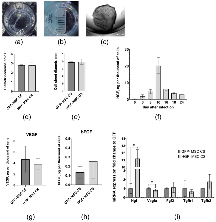
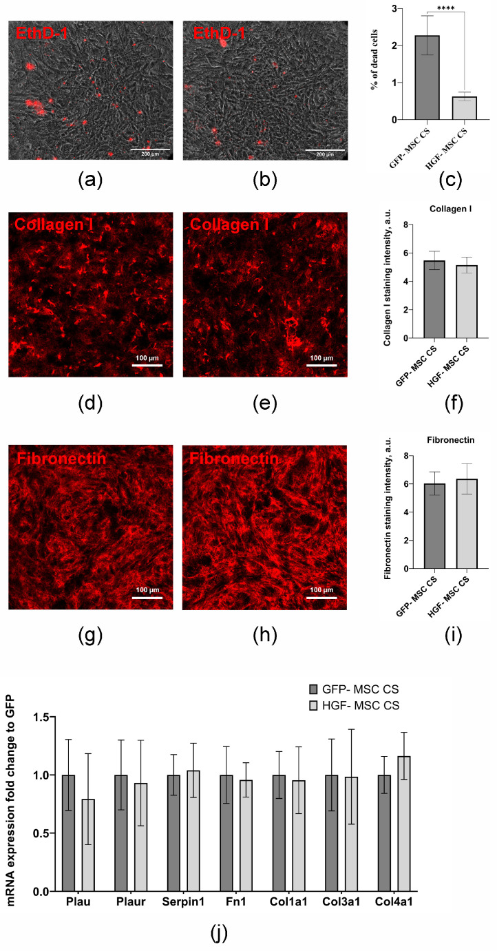
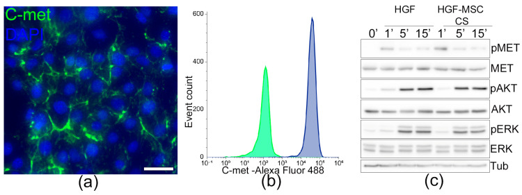
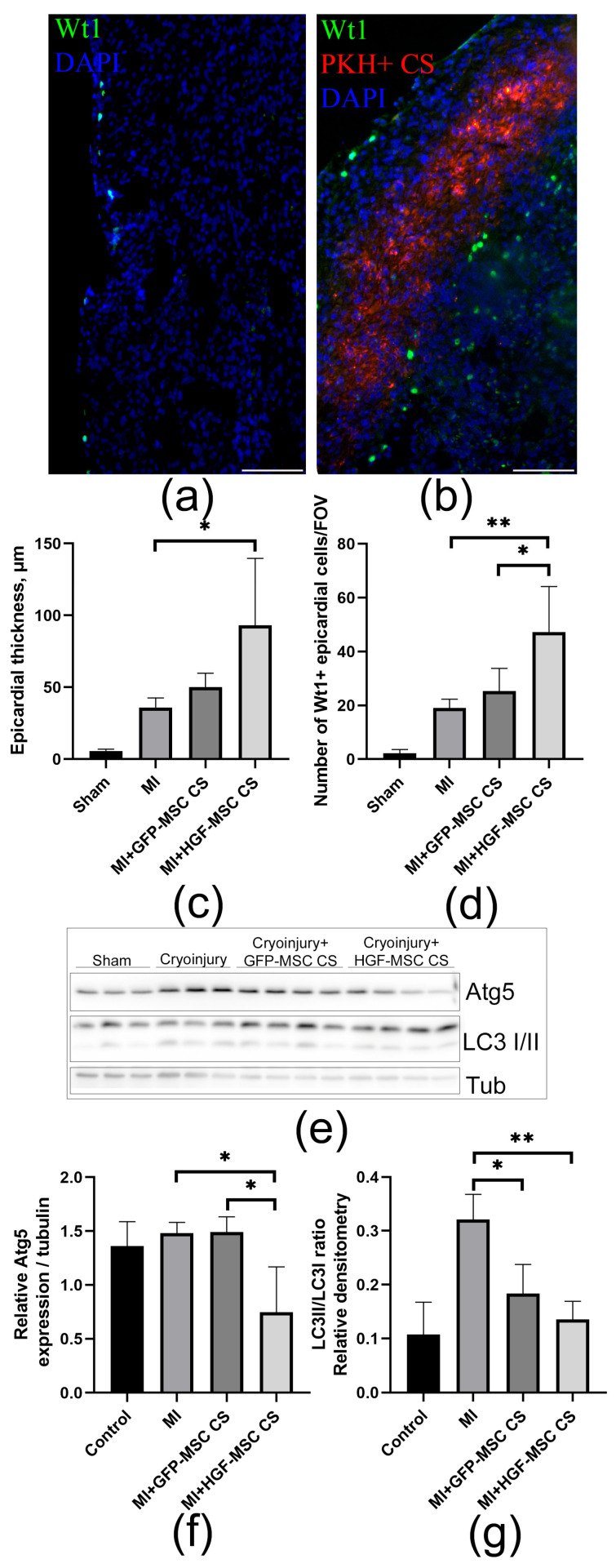
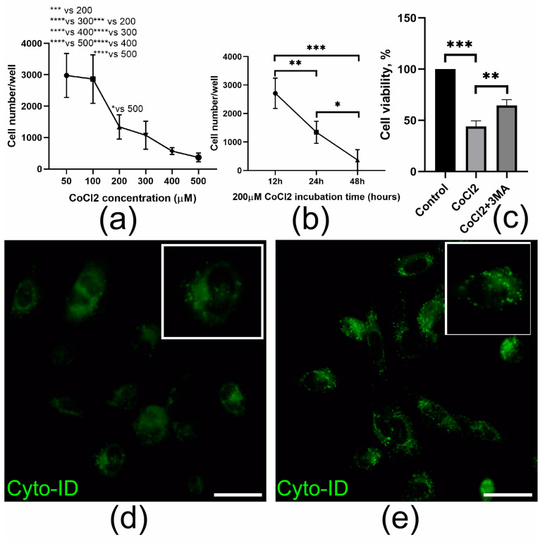
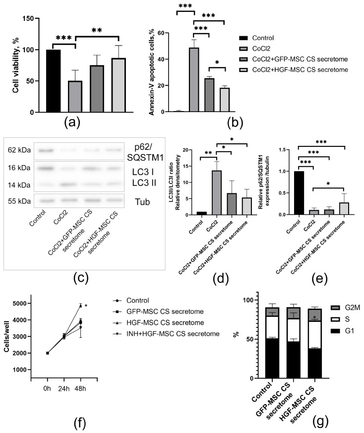
Epicardial mesothelial cells (EMCs), which form the epicardium, play a crucial role in cardiac homeostasis and repair. Upon damage, EMCs reactivate embryonic development programs, contributing to wound healing, progenitor cell amplification, and regulation of lymphangiogenesis, angiogenesis, and fibrosis. However, the mechanisms governing EMC activation and subsequent regulation remain poorly understood. We hypothesized that hepatocyte growth factor (HGF), a pleiotropic regulator of various cellular functions, could modulate EMC activity. To verify this hypothesis, we developed HGF-overexpressing mesenchymal stromal cell sheets (HGF-MSC CSs) and evaluated their effects on EMCs in vitro and in vivo. This study has revealed, for the first time, that EMCs express the c-Met (HGF receptor) on their surface and that both recombinant HGF and HGF-MSC CSs secretome cause c-Met phosphorylation, triggering downstream intracellular signaling. Our findings demonstrate that the HGF-MSC CSs secretome promotes cell survival under hypoxic conditions by modulating the level of autophagy. At the same time, HGF-MSC CSs stimulate EMC proliferation, promoting their amplification in the damage zone. These data demonstrate that HGF-MSC CSs can be considered a promising regulator of epicardial cell activity involved in heart repair after ischemic damage.

Keywords: HGF; cell sheets; epicardial cells; myocardial infarction.


Conflict of interest statement

The authors declare no conflicts of interest.

Figures

Figure 1
Figure 1
The characteristics of the CSs. The 72 h cell sheets were washed three times with Versene solution and then incubated in Versene solution for 3–5 min at 37 °C and 5% CO2. The representative images of the CSs: (a) partly detached CSs (brightfield image), (b) completely detached CSs (brightfield image), (c) completely detached CSs (phase-contrast image). The diameters of CSs from (c) were analyzed using the ImageJ software (v 1.54d, NIH, USA). (d) The average diameters of the detached GFP-MSC CSs and HGF-MSC CSs. (e) The average folds of the GFP-MSC CS and HGF-MSC CS contraction after detachment. (f) HGF production by HGF-MSC for 24 days evaluated by ELISA. (g) VEGF secretion by GFP-MSC CSs and HGF-MSC CSs for 48 h on day 8 after infection, as evaluated by ELISA. (h) bFGF secretion by GFP-MSC CSs and HGF-MSC CSs for 48 h on day 8 after infection evaluated by ELISA. (i) Analysis of growth factor genes. Total RNA of GFP-MSC and HGF-MSC was isolated on day 11 after infection, and specific mRNA levels were quantified by qRT PCR, as described in the Section 4. The data are presented as fold changes in the mRNA levels normalized to GFP-MSC “*”: p < 0.05, n ≥ 4.
Figure 2
Figure 2
Characteristics of CSs, whereby 72 h CSs were stained with LIVE/DEAD™ Viability/Cytotoxicity Kit. (a,b) % of dead cells in CS. Representative fluorescence images of CSs stained with ethidium homodimer-1: (a) GFP-MSC CSs and (b) HGF-MSC CSs. The percentage of dead cells was analyzed using ImageJ software (v 1.54d, NIH, USA) (c) Quantification of the percentage of the dead cells for GFP-MSC CSs and HGF-MSC CSs. (df) Analysis of collagen I deposition in the 72 h CSs. (d) GFP-MSC CSs and (e) HGF-MSC CSs were fixed with 4% formaldehyde, and stained with the anti-collagen I antibody (red). Images were taken using a Stellaris 5 confocal microscope (Leica, Wetzlar, Germany). (f) Quantification of the intensities of staining for collagen I using Image J software (v 1.54d, NIH, USA). (gi) Analysis of fibronectin deposition in the cell layer. (g) GFP-MSC CSs and (h) HGF-MSC CSs were fixed with 4% formaldehyde and stained with the anti-collagen I antibody (red). The images were taken using a Stellaris 5 confocal microscope (Leica, Germany). (i) Quantification of the intensities of staining for fibronectin using Image J software. (j) Analysis of extracellular matrix genes. Total RNA of GFP-MSC and HGF-MSC was isolated on day 11 after infection and specific mRNA levels were quantified by qRT PCR as described in the Section 4. The data are presented as fold changes in the mRNA levels normalized to GFP-MSC “****”: p < 0.001, n ≥ 4.
Figure 3
Figure 3
Mouse epicardial cells expressing the c-Met receptor and activating intracellular signaling upon exposure to HGF and HGF-MSC CSs secretome. (a) Immunocytochemical analysis of c-Met receptor expression (green) on the surface of the EMCs. The nuclei were counterstained with DAPI. Scale bar: 50 μM. (b) The results of the c-Met expression analysis in the EMCs were obtained using flow cytometry. Immunolabelling with c-Met antibodies (blue; control (green)) revealed its presence on the surface of more than 90% of the EMCs. (c) Phosphorylation of the c-Met receptor and Akt; ERK signaling pathways after exposure to recombinant HGF and HGF-MSC CSs secretome for 1, 5, and 15 min determined by western blot.
Figure 4
Figure 4
The transplantation of HGF-MSC CSs increases the number of Wt1+ EMCs in the damaged area and regulates the level of autophagy. (a,b) The representative images of heart tissue after cryoinjury (a) and cryoinjury+ HGF-MSC CSs transplantation (b) with antibodies against epicardial marker—Wt1 (green). The transplanted HGF-MSC CS (b) are labelled with PKH (red). The cell nuclei are stained with DAPI (blue). Scale bar: 50 mkm. (c,d) The thickness of the epicardial zone (c) and the number of Wt1+ EMCs (d) in the hearts of sham, cryoinjury, and cell sheet groups (cryoinjury after transplantation of HGF-MSC and GFP-MSC CSs) were calculated and presented in graphs. ANOVA; * p < 0.05, ** p < 0.01; n ≥ 4. (e) Atg5 and LC3 I/II expression were analyzed by western blot. The results are shown in the representative images. (f,g) Densitometric quantification of the Atg5 and LC3 II/I ratio in the epicardial cells of cryoinjured hearts (day 2 after surgery) of the control group and groups, which received a transplant of either GFP- or HGF-MSC CSs. Tubulin was used as loading control. All the data are expressed as mean ± standard deviation (SD) from at least three experiments. * p < 0.05, ** p < 0.01.
Figure 5
Figure 5
The CoCl2 treatment induces autophagosome formation and decreases EMCs survival. (a,b) Cell viability was analyzed using PrestoBlue assay after exposure to CoCl2 at the indicated doses (a) and time intervals (b). (c) Cell viability was analyzed using PrestoBlue assay in control (untreated), cells treated with CoCl2 (200 mkM) and CoCl2 (200 mkM) + 3MA(10 mkM). (d,e) Autophagic vacuoles in epicardial cells were visualized using Cyto-ID® in the untreated EMCs and cells after 12 h of exposure to CoCl2 (200 μM). Scale bar: 50 mkm. All the data are expressed as mean ± standard deviation (SD) from three or more replications. * p < 0.05, ** p < 0.01, *** p < 0.005 and **** p < 0.001.
Figure 6
Figure 6
The HGF-MSC CSs secretome protects EMCs from CoCl2-induced apoptosis and stimulates their proliferation. (a) Cell viability was analyzed using PrestoBlue assay in control (untreated) and cells treated with CoCl2 (200 mkM), CoCl2 (200 mkM) + GFP-MSC CSs secretome and CoCl2 (200 mkM) + HGF-MSC CSs secretome. The results are represented as relative viability (% of untreated control). (b) Apoptosis level was analyzed using Annexin-V/PI assay in control (untreated) and cells treated with CoCl2 (200 mkM), CoCl2 (200 mkM) + GFP-MSC CSs secretome and CoCl2 (200 mkM) + HGF-MSC CSs secretome. The results are represented as the presence of Annexin V+ cells relative to control. (c) The representative images of western blot analysis demonstrate the p62/SQSTM1 and LC3 I/II expression in the control (untreated) cells and cells treated with CoCl2 (200 mkM), CoCl2 (200 mkM) + GFP-MSC CSs secretome and CoCl2 (200 mkM) + HGF-MSC CSs secretome. (d,e) Densitometric quantification of the p62/SQSTM1 and LC3 II/I ratio in the control (untreated) and cells treated with CoCl2 (200 mkM), CoCl2 (200 mkM) + GFP-MSC CSs secretome and CoCl2 (200 mkM) + HGF-MSC CSs secretome. Tubulin was used as a loading control. (f) Growth curve of control and cells treated for 48 h with GFP-MSC CS and HGF-MSC CS secretomes. To determine the effect of HGF-MSC CSs secretome on cell proliferation, the c-Met inhibitor crizotinib (100 nM) was added to the culture medium. (g) The EMCs were incubated for 36 h in control or GFP-MSC CSs and HGF-MSC CSs secretomes. Cell cycle analysis was performed using flow cytometry. All the data are expressed as mean ± standard deviation (SD) from three or more replications. * p < 0.05, ** p < 0.01, and *** p < 0.005.

Similar articles

References

    1. Kumar Singh A., Kumar Jat R. Myocardial Infarction. Himal. J. Health Sci. 2022;6:16–32. doi: 10.22270/hjhs.v6i4.116. - DOI
    1. Al-Botaty B., El-Khoely A., Elsayed E.M., Eissa A.A. Insight into the Pathophysiology of Myocardial Infarction. Adv. Pharm. Res. 2022;6:223–237. doi: 10.21608/aprh.2022.155725.1188. - DOI
    1. Cao J., Poss K.D. The Epicardium as a Hub for Heart Regeneration. Nat. Rev. Cardiol. 2018;15:631–647. doi: 10.1038/s41569-018-0046-4. - DOI - PMC - PubMed
    1. Dergilev K.V., Komova A.V., Tsokolaeva Z.I., Beloglazova I.B., Parfyonova Y.V. Epicardium as a New Target for Regenerative Technologies in Cardiology. Genes Cells. 2020;15:33–40. doi: 10.23868/202004016. - DOI
    1. Li G., Yan Z., Han L., Wu S., Wang M., Qi A., Zhou Z., Wang N., Sun R., Zhou X. Research Progress on Epicardial Repair After Myocardial Injury. Cardiol. Rev. 2024 doi: 10.1097/CRD.0000000000000806. - DOI - PubMed

LinkOut - more resources