Review
. 2025 Jul 29;26(15):7328.
doi: 10.3390/ijms26157328.

Are Hippocampal Hypoperfusion and ATP Depletion Prime Movers in the Genesis of Alzheimer's Disease? A Review of Recent Pertinent Observations from Molecular Biology

Affiliations
Review

Are Hippocampal Hypoperfusion and ATP Depletion Prime Movers in the Genesis of Alzheimer's Disease? A Review of Recent Pertinent Observations from Molecular Biology

Valerie Walker. Int J Mol Sci. .

Abstract

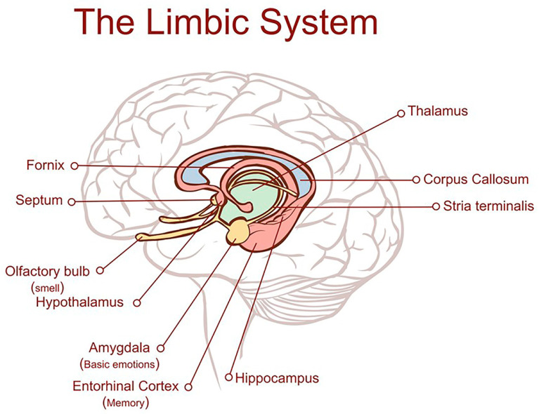
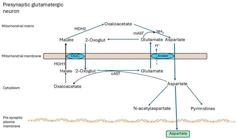
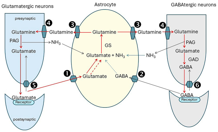
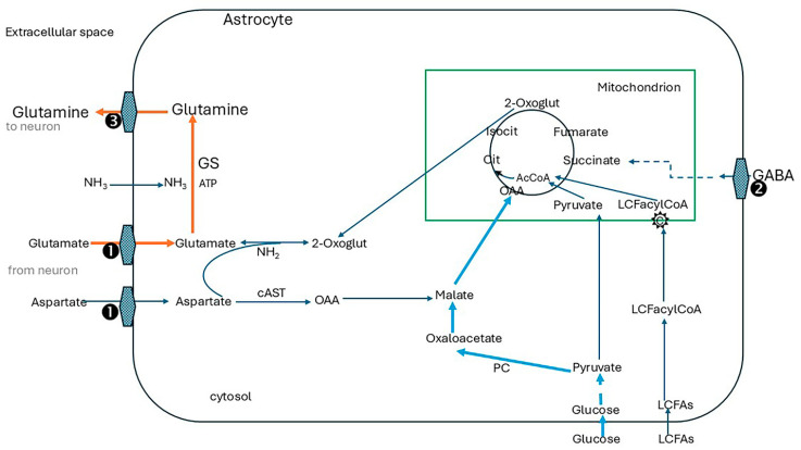
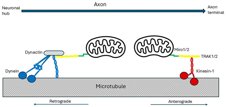
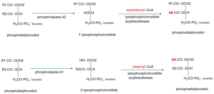
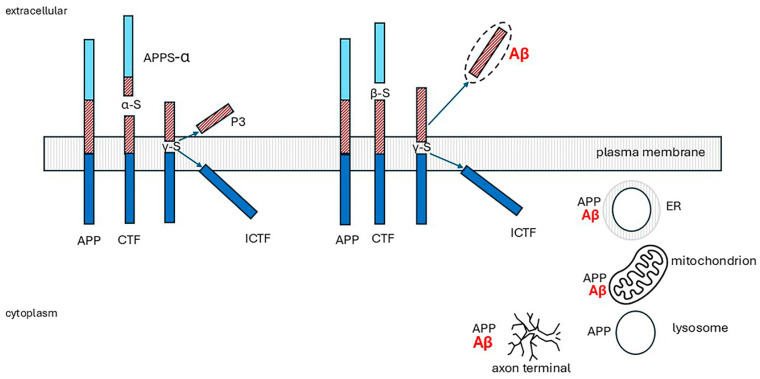
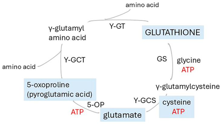
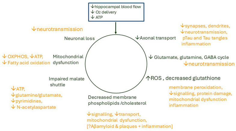
Alzheimer's dementia (AD) is a disease of the ageing brain. It begins in the hippocampal region with the epicentre in the entorhinal cortex, then gradually extends into adjacent brain areas involved in memory and cognition. The events which initiate the damage are unknown and under intense investigation. Localization to the hippocampus can now be explained by anatomical features of the blood vessels supplying this region. Blood supply and hence oxygen delivery to the area are jeopardized by poor flow through narrowed arteries. In genomic and metabolomic studies, the respiratory chain and mitochondrial pathways which generate ATP were leading pathways associated with AD. This review explores the notion that ATP depletion resulting from hippocampal hypoperfusion has a prime role in initiating damage. Sections cover sensing of ATP depletion and protective responses, vulnerable processes with very heavy ATP consumption (the malate shuttle, the glutamate/glutamine/GABA (γ-aminobutyric acid) cycle, and axonal transport), phospholipid disturbances and peroxidation by reactive oxygen species, hippocampal perfusion and the effects of hypertension, chronic hypoxia, and arterial vasospasm, and an overview of recent relevant genomic studies. The findings demonstrate strong scientific arguments for the proposal with increasing supportive evidence. These lines of enquiry should be pursued.

Keywords: ATP biosensors; axonal transport; cerebral arterial perfusion; glutamate/GABA/glutamine cycle; malate aspartate shuttle; membrane phospholipids; mitochondrial-derived peptides; vasospasm.


Conflict of interest statement

The author declares no conflicts of interest.

Figures

Figure 1
Figure 1
Location of the entorhinal cortex. The entorhinal cortex is part of the brain’s limbic system which controls emotional drives and memory formation. It is a collection of structures located deep within the brain which includes the hippocampal formation, amygdala, septal nuclei, cingulate cortex, entorhinal cortex, perirhinal cortex, and parahippocampal cortex. The last three cortical areas comprise different portions of the temporal lobe [6]; image ID HYTTNN hakan çorbac?/Alamy Stock Vector reproduced under license from Alamy Limited, Abingdon, UK, https://www.alamy.com. (accessed on 25 July 2025).
Figure 2
Figure 2
The malate-aspartate shuttle. NADH cannot be transported from the cytosol into mitochondria. To regenerate NAD from NADH produced during oxidative reactions in the cytosol, H+ from NADH is transported into mitochondria via the malate-aspartate shuttle (MAS). MAS requires two cytoplasmic enzymes, cAST and MDH1; two mitochondrial enzymes, mAST and mMDH2; and two carriers located in the inner mitochondrial membrane, the aspartate-glutamate carrier aralar (SCL25A12) and the 2-oxoglutarate carrier OGC (SLC25A11). (i) In the cytoplasm, MDH1 transfers reducing equivalents from NADH to oxaloacetate, producing malate; (ii) OGC transports malate into mitochondrion in exchange for 2-oxoglutarate (2-oxoglut); (iii) mMDH2 then oxidizes malate to oxaloacetate (OAA), generating NADH; (iv) mAST transfers NH2 from glutamate to OAA producing aspartate and 2-oxoglut; (v) aralar transports aspartate out into the cytoplasm in exchange for glutamate and H+ into the mitochondria; and (vi) finally, cAST transaminates 2-oxoglut forming OAA and glutamate, closing the cycle. After entry into mitochondria, electrons are supplied to the electron transport chain in the form of NADH for ATP production, and cytosolic NAD+ is regenerated [116,117,118]. The Glutamate/H+ symporter, SLCA22, may also contribute to the shuttle activity [18]. Abbreviations: MDH1 malate dehydrogenase 1, MDH2 malate dehydrogenase 2 (mitochondrial), mAST mitochondrial aspartate aminotransferase, alias GOT2 glutamic-oxaloacetic transaminase 2, mitochondrial, cAST cytoplasmic aspartate aminotransferase, alias GOT1 glutamic-oxaloacetic transaminase 1, 2-Oxoglut, 2-oxoglutarate, OGC 2-oxoglutarate carrier.
Figure 3
Figure 3
Overview of the glutamate/GABA/glutamine cycle. The GABA–glutamine–glutamate shuttles replenish the neurotransmitters, L-glutamate and GABA using glutamine generated in astrocytes. After release from glutamatergic neurons into the synaptic clefts, glutamate not bound to post-synaptic receptors is carried with Na+ and H+ into astrocytes by the glutamate–aspartate transporters SLC1A3 (solute carrier 1A3, EAAT1) and SLC1A2 (EAAT2), step ①. Similarly, unbound GABA released from GABAtergic neurons is carried into astrocytes with sodium and chloride by the GABA transporter SLC6A11 (GAT3), step ②, where it is converted to glutamate. Glutamine synthetase (GS) then catalyzes the formation of glutamine from glutamate and ammonia in an ATP-dependent reaction. Glutamine is exported with sodium into the extracellular space by Na+-amino acid cotransporters, SLC38A1 (SNAT1) and SLC38A2 (SNAT2), and Na+-amino acid cotransporters-H+ antiporters, SLC38A3 (SNAT3) and SLC38A5 (SNAT5), step ③, and then imported into neurons by one or more SNAT transporters: SNAT1, 2 and SNAT7 [SLC38A7]), step ④. In glutamatergic neurons glutamine is hydrolysed by PAG (phosphate-activated glutaminase). In GABAtergic neurons. glutamine is dehydrogenated by GDH (glutamate dehydrogenase), producing GABA. The neurotransmitters are then packaged into vesicles, transported to the synapses, and released with neuronal stimulation. Most neurotransmitters not bound to post-synaptic receptors are recycled via astrocytes as described above. A fraction is transferred back into neurons: glutamate by SLC1A3 ⑤ and GABA by SLC6A1 (GAT1) ⑥. Mitochondrial glutamate/H+ symporter SLCA22 probably makes a significant contribution to the cycle [18]. Abbreviations: Solute carrier family members (SLC): SLC1A3 aliases excitatory amino acid transporter 1 (EAAT1), glutamate-aspartate transporter (GLAST), SLC1A2, aliases EAAT2, glutamate transporter 1 (GLT1), SLC6A1 (alias GABA transporter 1 (GAT1), SLC6A11, alias GABA transporter 3 (GAT3); Na+-amino acid cotransporters: SLC38A1 (SNAT1) and SLC38A2 (SNAT2); Na+-amino acid cotransporters-H+ antiporters, SLC38A3 (SNAT3), SLC38A5 (SNAT5), and SLC38A7 (SNAT7). Red lines depict the major pathways, grey lines additional routes. Solid lines indicate direct enzyme conversions; dotted lines depict production from TCA cycle intermediates.
Figure 4
Figure 4
Astrocytes are central to neurotransmitter turnover of glutamatergic neurones. Glutamine is replenished in astrocytes by transamination of 2-oxoglutarate drawn from the TCA cycle, producing glutamate which GS then converts to glutamine. TCA cycle function is maintained largely by carboxylation of pyruvate (provided by glucose) by cytosolic pyruvate carboxylase forming oxaloacetate. This enters the cycle after hydrogenation to malate and transport into mitochondria. Oxaloacetate is also produced from aspartate generated by the malate-aspartate shuttle and makes an essential contribution to anaplerosis. Further anaplerotic support is provided by reuptake of GABA into astrocytes and its conversion to succinate. ① SLC1A3, alias excitatory amino acid transporter 1 (EAAT1; GLAST), SLC1A2, alias excitatory amino acid transporter 2 (EAAT2, GLT1), ② SLC6A11 (GAT3), ③ Na+-amino acid cotransporters, SLC38A1 (SNAT1) and SLC38A2 (SNAT2), and Na+-amino acid cotransporters-H+ antiporters, SLC38A3 (SNAT3) and SLC38A5 (SNAT5). cAST cytoplasmic aspartate aminotransferase (alias GOT1 glutamic-oxaloacetic transaminase 1, cytoplasmic), GS glutamine synthetase, PC pyruvate carboxylase, OAA oxaloacetate, 2-oxoglut 2-oxoglutarate, cit citrate, isocit isocitrate, AcCoA acetyl CoA, LCFA long-chain fatty acids, LCFacylCoA, and C carnitine shuttle. Red lines, main route for glutamine synthess; solid blue lines, main anaplerotic pathway from glucose via pyruvate carboxylase; blue dotted lines anaplerosis from GABA via conversion to succinate.
Figure 5
Figure 5
Axonal transport of mitochondria. For transport down axons, mitochondria attach to an adaptor which, in turn, links them to a motor bound to microtubules. Miro proteins (green) anchored to the outer mitochondrial membrane bind to trafficking kinesin protein adaptor complex adaptors, TRAK1 or TRAK 2 (yellow). For anterograde travel to the nerve terminals, TRAK1/2 binds to the kinesin-1 motor (red). This has two heavy and two light chains. The heavy chains dimerize and their head domains in alternation bind and then detach from myosin, consuming ATP, and ‘walk’ down the microtubules. Their C-terminals bind to the cargo-carrying light chains. The multimeric complex dynein and its activator dynactin (blue) transport damaged mitochondria from the axon terminals to the cell body for elimination by mitophagy [178,179,186,187].
Figure 6
Figure 6
Remodelling of phosphatidylinositides by the Lands cycle. The phosphatidylinositides (PIs) synthesized de novo have a miscellany of fatty acyl groups. Phospholipase A1 or A2 removes the acyl chains at sn-1 or sn-2, respectively, leaving a lysophosphoinositide. The new, required acyl groups (shown in red) are transferred to this from stearoyl-CoA or arachidonyl-CoA. PIs incorporated into glycosylphosphatidylinositol (GPI)-anchored proteins are remodelled by the same process, but with replacement of flexible unsaturated fatty acyl chains at sn-2 with rigid stearate chains transferred from stearoyl-CoA [127,227]. Polyunsaturated acyl groups located at the sn-2 position have a rapid turnover. Synthesis, remodelling, and recycling of phospholipids are highly active processes with high ATP consumption [228,229]. AA arachidonic acid, SA stearic acid.
Figure 7
Figure 7
Processing of amyloid precursor protein (APP), not drawn to scale. Full length APP has three components: a large extracellular domain (light blue and brown) representing around 85% of protein mass, a short transmembrane sequence (brown and dark blue) and a small cytoplasmic domain (dark blue). APP is first cleaved extracellularly. In the non-amyloidogenic pathway (left), α-secretase (α-s) releases a soluble peptide, APPs-α. The 83-residue membrane-bound fragment is then cleaved by γ-secretase (γ-s) in its transmembrane domain releasing a soluble N-terminal fragment, P3 peptide and an intracellular C-terminal fragment (ICTF). In the amyloidogenic pathway (right), β-secretase (β-s, β-APP-cleaving enzyme 1, BACE) cleaves APP at a more proximal site, releasing an N-terminal peptide (light blue). The 99-residue membrane-bound fragment is then cleaved by γ-secretase, possibly at more than one site. ICTF is released into the cytoplasm; Aβ peptides with 39 to 42 residues are discharged extracellularly. They may aggregate, oligomerize, and form fibrils [34,49,249].
Figure 8
Figure 8
The γ-glutamyl cycle 5-oxoproline (pyroglutamic acid) is an intermediate in glutathione metabolism. The tripeptide glutathione is synthesized and degraded in the γ-glutamyl cycle. It is synthesized from glutamate by the sequential actions of γ-glutamylcysteine synthetase (γ-GCS) and glutathione synthetase (GS). γ-glutamine transpeptidase (γ-GT) initiates breakdown by transferring the γ-glutamyl group to amino acid acceptors. γ-glutamylcyclotransferase (γ-GCT) forms 5-oxoproline and releases the amino acids. Notably, 5-oxoprolinase (5-OP) converts 5-oxoproline to glutamate. A dipeptidase (not shown) splits the cysteinylglycine moiety of glutathione into cysteine and glycine. The cycle has heavy ATP consumption.
Figure 9
Figure 9
The impact of low hippocampal blood flow on neuronal activities. It is proposed that reduction in blood flow, causing poor perfusion of the hippocampus, reduces oxygen delivery to the nerve cells, and ATP synthesis falls. This disrupts essential neuronal activities (shown in dark green) precipitating a cascade of biochemical disturbances (shown in orange). Repeated episodes cause cumulative damage and inflammation which slowly spreads further into the brain and impacts memory and learning. ↓ decreased; ↑ increased; ? speculated may be implicated in Aβ and plaque formation.

Similar articles

  • Prescription of Controlled Substances: Benefits and Risks.
    Preuss CV, Kalava A, King KC. Preuss CV, et al. 2025 Jul 6. In: StatPearls [Internet]. Treasure Island (FL): StatPearls Publishing; 2025 Jan–. 2025 Jul 6. In: StatPearls [Internet]. Treasure Island (FL): StatPearls Publishing; 2025 Jan–. PMID: 30726003 Free Books & Documents.
  • Short-Term Memory Impairment.
    Cascella M, Al Khalili Y. Cascella M, et al. 2024 Jun 8. In: StatPearls [Internet]. Treasure Island (FL): StatPearls Publishing; 2025 Jan–. 2024 Jun 8. In: StatPearls [Internet]. Treasure Island (FL): StatPearls Publishing; 2025 Jan–. PMID: 31424720 Free Books & Documents.
  • Selegiline for Alzheimer's disease.
    Birks J, Flicker L. Birks J, et al. Cochrane Database Syst Rev. 2003;(1):CD000442. doi: 10.1002/14651858.CD000442. Cochrane Database Syst Rev. 2003. PMID: 12535396
  • [Volume and health outcomes: evidence from systematic reviews and from evaluation of Italian hospital data].
    Amato L, Colais P, Davoli M, Ferroni E, Fusco D, Minozzi S, Moirano F, Sciattella P, Vecchi S, Ventura M, Perucci CA. Amato L, et al. Epidemiol Prev. 2013 Mar-Jun;37(2-3 Suppl 2):1-100. Epidemiol Prev. 2013. PMID: 23851286 Italian.
  • MarkVCID cerebral small vessel consortium: I. Enrollment, clinical, fluid protocols.
    Wilcock D, Jicha G, Blacker D, Albert MS, D'Orazio LM, Elahi FM, Fornage M, Hinman JD, Knoefel J, Kramer J, Kryscio RJ, Lamar M, Moghekar A, Prestopnik J, Ringman JM, Rosenberg G, Sagare A, Satizabal CL, Schneider J, Seshadri S, Sur S, Tracy RP, Yasar S, Williams V, Singh H, Mazina L, Helmer KG, Corriveau RA, Schwab K, Kivisäkk P, Greenberg SM; MarkVCID Consortium. Wilcock D, et al. Alzheimers Dement. 2021 Apr;17(4):704-715. doi: 10.1002/alz.12215. Epub 2021 Jan 21. Alzheimers Dement. 2021. PMID: 33480172 Free PMC article.

References

    1. Gómez-Isla T., Price J.L., McKeel D.W., Jr., Morris J.C., Growdon J.H., Hyman B.T. Profound loss of layer II entorhinal cortex neurons occurs in very mild Alzheimer’s disease. J. Neurosci. 1996;16:4491–4500. doi: 10.1523/JNEUROSCI.16-14-04491.1996. - DOI - PMC - PubMed
    1. Morgan G.R., Carlyle B.C. Interrogation of the human cortical peptidome uncovers cell-type specific signatures of cognitive resilience against Alzheimer’s disease. Sci. Rep. 2024;14:7161. doi: 10.1038/s41598-024-57104-z. - DOI - PMC - PubMed
    1. Thangavel R., Kempuraj D., Stolmeier D., Anantharam P., Khan M., Zaheer A. Glia maturation factor expression in entorhinal cortex of Alzheimer’s disease brain. Neurochem. Res. 2013;38:1777–1784. doi: 10.1007/s11064-013-1080-6. - DOI - PMC - PubMed
    1. Kapadia A., Billimoria K., Desai P., Grist J.T., Heyn C., Maralani P., Symons S., Zaccagna F. Hypoperfusion Precedes Tau Deposition in the Entorhinal Cortex: A Retrospective Evaluation of ADNI-2 Data. J. Clin. Neurol. 2023;19:131–137. doi: 10.3988/jcn.2022.0088. - DOI - PMC - PubMed
    1. Kahn I., Andrews-Hanna J.R., Vincent J.L., Snyder A.Z., Buckner R.L. Distinct cortical anatomy linked to subregions of the medial temporal lobe revealed by intrinsic functional connectivity. J. Neurophysiol. 2008;100:129–139. doi: 10.1152/jn.00077.2008. - DOI - PMC - PubMed

LinkOut - more resources