

Sensitivity of Diffusion Tensor Imaging for Assessing Injury Severity in a Rat Model of Isolated Diffuse Axonal Injury: Comparison with Histology and Neurological Assessment
- PMID: 40806467
- PMCID: PMC12347101
- DOI: 10.3390/ijms26157333
Sensitivity of Diffusion Tensor Imaging for Assessing Injury Severity in a Rat Model of Isolated Diffuse Axonal Injury: Comparison with Histology and Neurological Assessment
Abstract
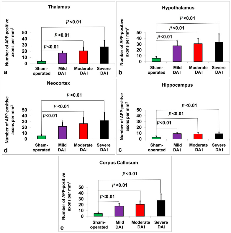
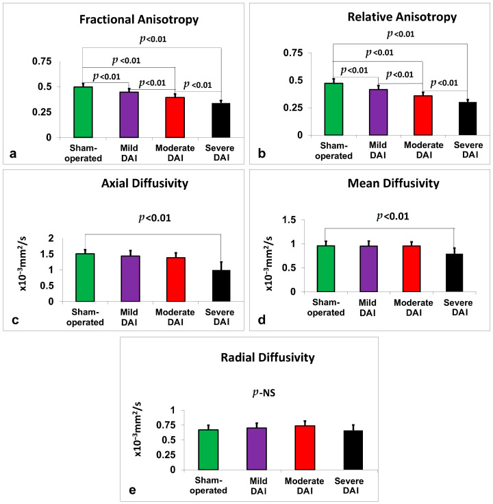
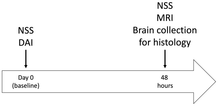
Diffuse axonal brain injury (DAI) is a common, debilitating consequence of traumatic brain injury, yet its detection and severity grading remain challenging in clinical and experimental settings. This study evaluated the sensitivity of diffusion tensor imaging (DTI), histology, and neurological severity scoring (NSS) in assessing injury severity in a rat model of isolated DAI. A rotational injury model induced mild, moderate, or severe DAI in male and female rats. Neurological deficits were assessed 48 h after injury via NSS. Magnetic resonance imaging, including DTI metrics, such as fractional anisotropy (FA), relative anisotropy (RA), axial diffusivity (AD), mean diffusivity (MD), and radial diffusivity (RD), was performed prior to tissue collection. Histological analysis used beta amyloid precursor protein immunohistochemistry. Sensitivity and variability of each method were compared across brain regions and the whole brain. Histology was the most sensitive method, requiring very small groups to detect differences. Anisotropy-based MRI metrics, especially whole-brain FA and RA, showed strong correlations with histology and NSS and demonstrated high sensitivity with low variability. NSS identified injury but required larger group sizes. Diffusivity-based MRI metrics, particularly RD, were less sensitive and more variable. Whole-brain FA and RA were the most sensitive MRI measures of DAI severity and were comparable to histology in moderate and severe groups. These findings support combining NSS and anisotropy-based DTI for non-terminal DAI assessment in preclinical studies.
Keywords: axonal injury; diffusion tensor imaging; histology; magnetic resonance imaging; neurological assessment; traumatic brain injury.
Conflict of interest statement
The authors declare no conflict of interest.
Figures
Similar articles
-
Translating state-of-the-art spinal cord MRI techniques to clinical use: A systematic review of clinical studies utilizing DTI, MT, MWF, MRS, and fMRI.Neuroimage Clin. 2015 Dec 4;10:192-238. doi: 10.1016/j.nicl.2015.11.019. eCollection 2016. Neuroimage Clin. 2015. PMID: 26862478 Free PMC article.
-
Exploring IVIM-DKI and DKI for Assessing Microvascular and Microstructural Changes After Traumatic Brain Injury.NMR Biomed. 2025 Sep;38(9):e70110. doi: 10.1002/nbm.70110. NMR Biomed. 2025. PMID: 40751360
-
Noninvasive assessment of glymphatic dysfunction and IDH mutation in glioma with DTI-ALPS and DTI metrics.Quant Imaging Med Surg. 2025 Jun 6;15(6):5007-5022. doi: 10.21037/qims-2024-2710. Epub 2025 May 19. Quant Imaging Med Surg. 2025. PMID: 40606349 Free PMC article.
-
A systematic review and data synthesis of longitudinal changes in white matter integrity after mild traumatic brain injury assessed by diffusion tensor imaging in adults.Eur J Radiol. 2022 Feb;147:110117. doi: 10.1016/j.ejrad.2021.110117. Epub 2021 Dec 23. Eur J Radiol. 2022. PMID: 34973540
-
Prescription of Controlled Substances: Benefits and Risks.2025 Jul 6. In: StatPearls [Internet]. Treasure Island (FL): StatPearls Publishing; 2025 Jan–. 2025 Jul 6. In: StatPearls [Internet]. Treasure Island (FL): StatPearls Publishing; 2025 Jan–. PMID: 30726003 Free Books & Documents.
References
-
- Oleshko A., Gruenbaum B.F., Zvenigorodsky V., Shelef I., Negev S., Merzlikin I., Melamed I., Zlotnik A., Frenkel A., Boyko M. The role of isolated diffuse axonal brain injury on post-traumatic depressive-and anxiety-like behavior in rats. Transl. Psychiatry. 2025;15:113. doi: 10.1038/s41398-025-03333-3. - DOI - PMC - PubMed
-
- Drieu A., Lanquetin A., Prunotto P., Gulhan Z., Pédron S., Vegliante G., Tolomeo D., Serrière S., Vercouillie J., Galineau L. Persistent neuroinflammation and behavioural deficits after single mild traumatic brain injury. J. Cereb. Blood Flow Metab. 2022;42:2216–2229. doi: 10.1177/0271678X221119288. - DOI - PMC - PubMed
LinkOut - more resources
Full Text Sources
Miscellaneous
