. 2025 Jul 30;26(15):7361.
doi: 10.3390/ijms26157361.

Direct and Indirect Downstream Pathways That Regulate Repulsive Guidance Effects of FGF3 on Developing Thalamocortical Axons

Affiliations

Direct and Indirect Downstream Pathways That Regulate Repulsive Guidance Effects of FGF3 on Developing Thalamocortical Axons

Kejuan Li et al. Int J Mol Sci. .

Abstract

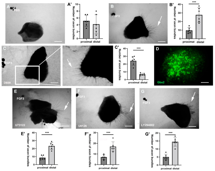
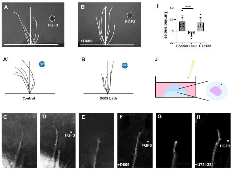
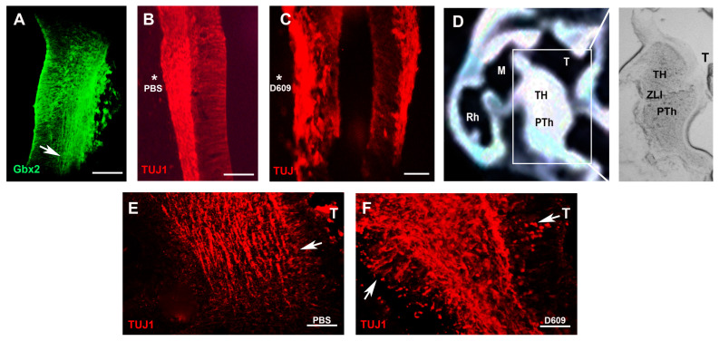
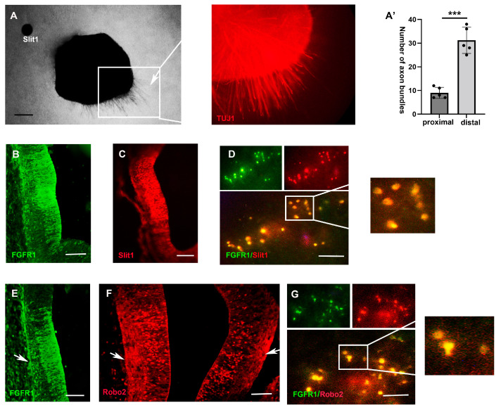
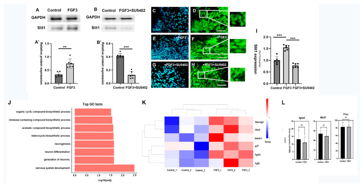
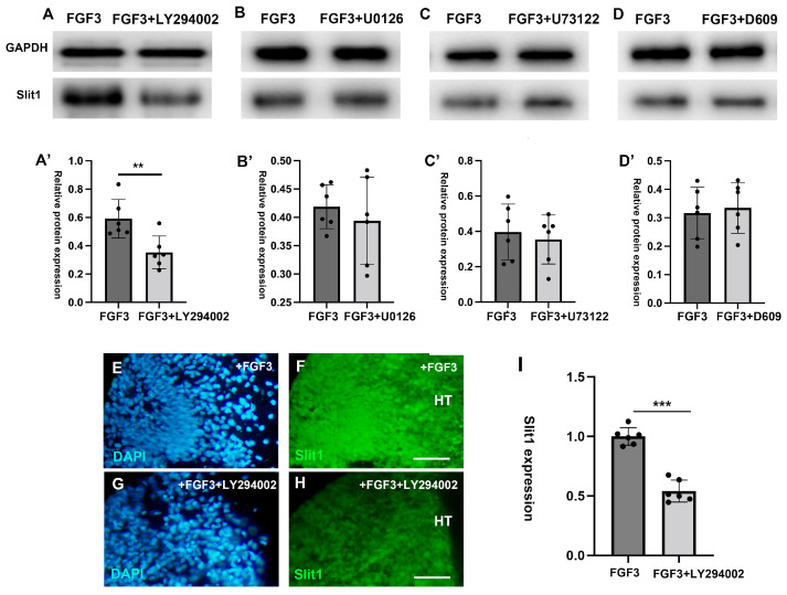
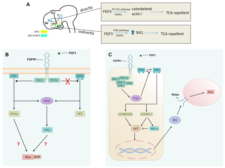
The thalamus is an important sensory relay station. It integrates all somatic sensory pathways (excluding olfaction) and transmits information through thalamic relay neurons before projecting to the cerebral cortex via thalamocortical axons (TCAs). Emerging evidence has shown that FGF3, a member of the morphogen family, is an axon guidance molecule that repels TCAs away from the hypothalamus and into the internal capsule so that they subsequently reach different regions of the cortex. However, current studies on FGF-mediated axon guidance predominantly focus on phenomenological observations, with limited exploration of the underlying molecular mechanisms. To address this gap, we investigated both direct and indirect downstream signaling pathways mediating FGF3-dependent chemorepulsion of TCAs at later developmental stages. Firstly, we used pharmacological inhibitors to identify the signaling cascade(s) responsible for FGF3-triggered direct chemorepulsion of TCAs, in vitro and in vivo. Our results demonstrate that the PC-PLC pathway is required for FGF3 to directly stimulate the asymmetrical repellent growth of developing TCAs. Then, we found the FGF3-mediated repulsion can be indirectly induced by Slit1 because the addition of FGF3 in the culture media induced an increase in Slit1 expression in the diencephalon. Furthermore, by using downstream inhibitors, we found that the indirect repulsive effect of FGF3 is mediated through the PI3K downstream pathway of FGFR1.

Keywords: FGF3; axon guidance; downstream pathways; thalamocortical axons; thalamus.


Conflict of interest statement

The authors have declared no conflicts of interest.

Figures

Figure 1
Figure 1
Pharmacological inhibition of PC-PLC pathway affects the TCA-repulsive effect of FGF3. Thalamic explants were co-cultured with FGF3 beads (500 ng/mL), and inhibitors of different pathways were added to the culture medium (n ≥ 6 in each group). (AC’) Compared with the control groups (A,A’: negative PBS control; B,B’: positive FGF3 control), when cultured with PC-PLC inhibitor D609 (10 µM), FGF3 beads lose their ability to repel TCA axons, even exhibiting attractive effects, since more thalamic axons (arrow) were observed on the proximal side (towards FGF3 beads) (C,C’). (D) The expression of glutamatergic marker Gbx2 in thalamic explants. (EG’) The inhibitors of U73122, U0126, and LY294002, however, show little effects on the TCA axonal rejection of FGF3. Scale bars (AC,EG) = 80 µm; ***: p < 0.001; Scale bar (D) = 40 µm.
Figure 2
Figure 2
FGF3 induces the repulsion of TCA growth cones by PC-PLC signaling pathway. (AB’) A FGF3 bead placed 25–50 µm away from the growth cone extending from a 24 h old thalamic explants for 3 h, with or without inhibitor (such as D609) in the media. (C,D) Phase micrographs of a representative growth cones in control media at t = 0 min (C), and after 3 h in the presence of an FGF3 gradient (D). (E,F) Trajectories of growth cones at t = 0 min (E), and at t = 3 h in 10 µM D609-supplemented media (F). (G,H) Growth cones prior to applying the FGF3 beads (t = 0 min) in control media (G), and growth cones 3 h after continuous exposure to FGF3 in the presence of 10 µM U73122 (H). (I) Differences in mean turning angles for distinct groups. (J) A schematic diagram showing the placement of FGF3 beads to the growth cones. ***: p < 0.001; asterisk: the implanation of FGF3 beads; scale bars = 25 µm.
Figure 3
Figure 3
The PC-PLC pathway is necessary for TCA extension in vivo. (A) The expression of thalamic glutamatergic marker Gbx2 in a transverse thalamic section at E5. (B) In a control bead implant (asterisk), transverse sections show thalamic pioneer projections at E5. (C) After a D609 bead implantation (asterisk) for 2 days, pioneer projections in the thalamus appear to be disorganized at E5. (D) Schematic showing a sagittal view of the E5 forebrain; the right-hand panel shows a high-power view of the boxed region. (E) PBS controls show that thalamic TUJ1+ axons (arrow) project towards the E5 prethalamus in a sagittal view. (F) With the addition of D609, the extension of thalamic axons is chaotic, and some axons (arrows) are outside the axonal bundle. TH, thalamus; PTh, prethalamus; M, midbrain; Rh, rhombencephalon; T, telencephalon; ZLI, zona limitans intrathalamica. Scale bars (AC) = 50 µm; scale bars (E,F) = 25 µm.
Figure 4
Figure 4
Expression patterns of Slit1 and downstream Robo2/FGFR1 genes in the E5 chick diencephalon. (A,A’) Slit1 beads show repellent effects on thalamic axons, and the right-hand panel shows TUJ1 expressions on thalamic axons of the boxed area. There are significantly more axon bundles from distal faces of thalamic explants (away from Slit1 beads) (n = 6 explants, *** p < 0.0001). (B,C) Immunofluorescence shows FGFR1 and Slit1 expressions in the diencephalon. (D) Double-labelled analyses showing co-expressions (orange) of Robo2 and FGFR1. (E,F) Comparison of Robo2 (E) and FGFR1 (F) expressions in the developing diencephalon and in TCAs (arrows). (G) Double-labelled analyses showing co-expressions (orange) of Robo2 and FGFR1. ***: p <0.001; scale bars (AC,E,F) = 50 µm; scale bars (D,G) = 40 µm.
Figure 5
Figure 5
FGF3 enhances the expression of repellent molecule Slit1. (A,A’) Western blot analyses showed that, compared with blank controls, FGF3 (300 ng/mL) in the culture media significantly increased the expression of Slit1 (n = 6). (B,B’) In contrast, the FGFR inhibitor SU5402 (20 µM) reduced the expression of the repellent factor Slit1 (n = 6). (CH) Enhanced immunofluorescence staining of DAPI (blue) and Slit1 (green) shows FGF3 upregulates Slit1 expression: explants of E4 diencephalon were cultured either for two days alone (C,D), with FGF3 (E,F), or with FGF3+SU5402 in the media (G,H). (I) Quantification of fluorescent signal from immuno-labelled Slit1 per volume of the thalamic explant. (J) Top GO items of RNA-seq were shown according to the gene counts. (K) Heatmap visualizes mRNA expression patterns of relative genes across different groups (controls and FGF3-treated). (L) Bar graphs for specific genes at mRNA transcription level (Vglut2, Mki67, Pcna), comparing their expression between controls and FGF3-treated explants. ns: Not-significant; FPKM, fragments per kilobase of transcript per million mapped reads; GO, gene ontology; **: p < 0.01; ***: p < 0.001; Scale bars = 40 µm.
Figure 6
Figure 6
PI3K downstream signaling is required for Slit1 expression in the diencephalon. Western blot analyses of Slit1 expression in the diencephalic explants on the second day post the addition of different pathway inhibitors (A,A’). Compared with FGF3 positive controls, PI3K pathway inhibitor LY294002 significantly reduced the expression of slit1 (BD’). However, the other pathway inhibitors U0126, D609 and U73122 had no significant effect on the expression of slit1 (EH). Immunofluorescence staining of DAPI (blue) and Slit1 (green) in the diencephalic explants on the second day post the addition of FGF3 (E,F), or post the addition of FGF3+LY294002 (G,H). (I) Quantification analyses of Slit1 per volume of the thalamic explant. **: p < 0.01; ***: p < 0.001; scale bars = 80 µm; HT: hypothalamus.
Figure 7
Figure 7
Working model of FGF3 on the guidance of TCAs in the thalamus. (A) FGF3 acts directly to regulate the turning of dorsal TCAs, which may be achieved by regulating the reorganization of PC-PLC downstream cytoskeletal actin. FGF3 is enriched in the ventral hypothalamus, potentially affecting expression levels of Slit1 activity by the PI3K pathway, and thus indirectly regulates the pathfinding of TCAs. (B) Schematic details the PC-PLC pathway activated by FGFR1 and FGF3, depicting components like PLC, DAG, IP3, PKC, and unresolved links to Rho and GTP. (C) Schematic depicts the indirect PI3K pathway, where FGFR1/FGF3 trigger signaling through molecules like SHP2, GRB2, etc., ultimately affecting Slit1, and cytoskeleton regulatory protein Rac. TH: thalamus; PT: prethalamus; HT: hypothalamus; T: telencephalon; ZLI: zona limitans intrathalamica; FGF: fibroblast growth factor; FGFR: fibroblast growth factor receptor; PC: phosphatidylcholine; PLC: phospholipase C; PLCγ: phospholipase C gamma; PI3K: phosphoinositide 3 kinase; PIP2: phosphatidylinositol 4,5-bisphosphate; DAG: diacylglycerol; IP3: inositol 1,4,5-trisphosphate; PCho: phosphocholine; PKC: protein kinase C; SHP2: Src homology 2 domain-containing protein tyrosine phosphatase 2; FRS2α: fibroblast growth factor receptor substrate 2 alpha; GRB2: growth factor receptor-bound protein 2; CRKL: CRK-like proto-oncogene; GAB1: Grb2-associated binding protein 1; cCOMPASS: catalytically active COMPASS; mTORC2: mammalian target of rapamycin complex 2; AKT: protein kinase B; Robo: Roundabout; red ×: the assumed pathway does not exist; red ?: probable pathways.

Similar articles

References

    1. Stoeckli E.T. Understanding axon guidance: Are we nearly there yet? Development. 2018;145:dev151415. doi: 10.1242/dev.151415. - DOI - PubMed
    1. Desbois M., Grill B. Molecular regulation of axon termination in mechanosensory neurons. Development. 2024;151:dev202945. doi: 10.1242/dev.202945. - DOI - PMC - PubMed
    1. Zhang W., Luo P., Liu X., Cheng R., Zhang S., Qian X., Liu F. Roles of Fibroblast Growth Factors in the Axon Guidance. Int. J. Mol. Sci. 2023;24:10292. doi: 10.3390/ijms241210292. - DOI - PMC - PubMed
    1. Tome D., Dias M.S., Correia J., Almeida R.D. Fibroblast growth factor signaling in axons: From development to disease. Cell Commun. Signal. 2023;21:290. doi: 10.1186/s12964-023-01284-0. - DOI - PMC - PubMed
    1. Short C.A., Onesto M.M., Rempel S.K., Catlett T.S., Gomez T.M. Familiar growth factors have diverse roles in neural network assembly. Curr. Opin. Neurobiol. 2021;66:233–239. doi: 10.1016/j.conb.2020.12.016. - DOI - PMC - PubMed

LinkOut - more resources