. 2025 Jul 30;26(15):7372.
doi: 10.3390/ijms26157372.

Circadian Light Manipulation and Melatonin Supplementation Enhance Morphine Antinociception in a Neuropathic Pain Rat Model

Affiliations

Circadian Light Manipulation and Melatonin Supplementation Enhance Morphine Antinociception in a Neuropathic Pain Rat Model

Nian-Cih Huang et al. Int J Mol Sci. .

Abstract

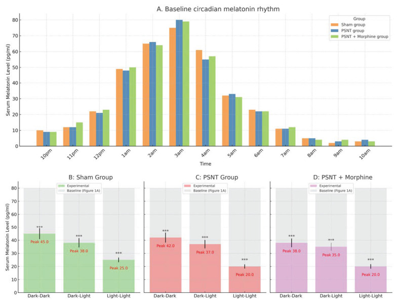
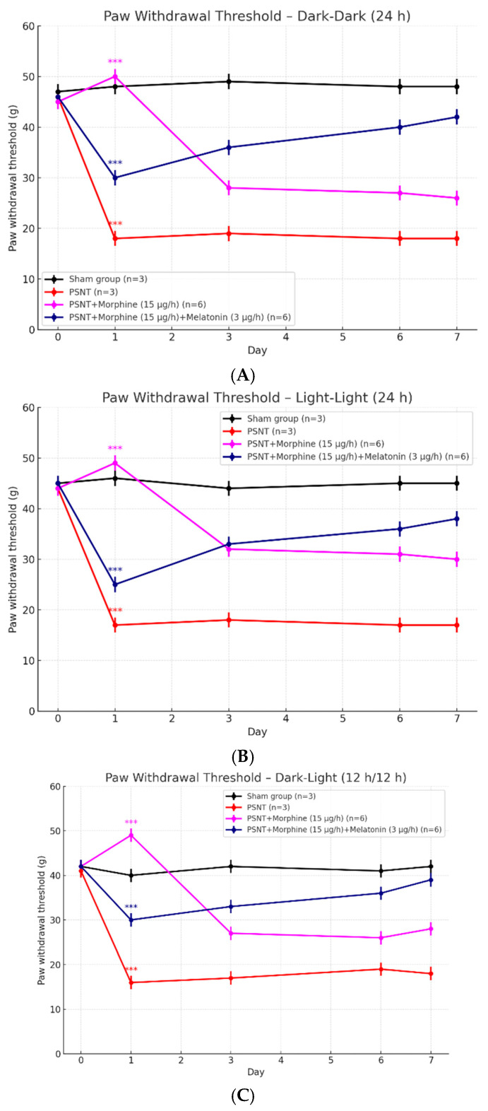
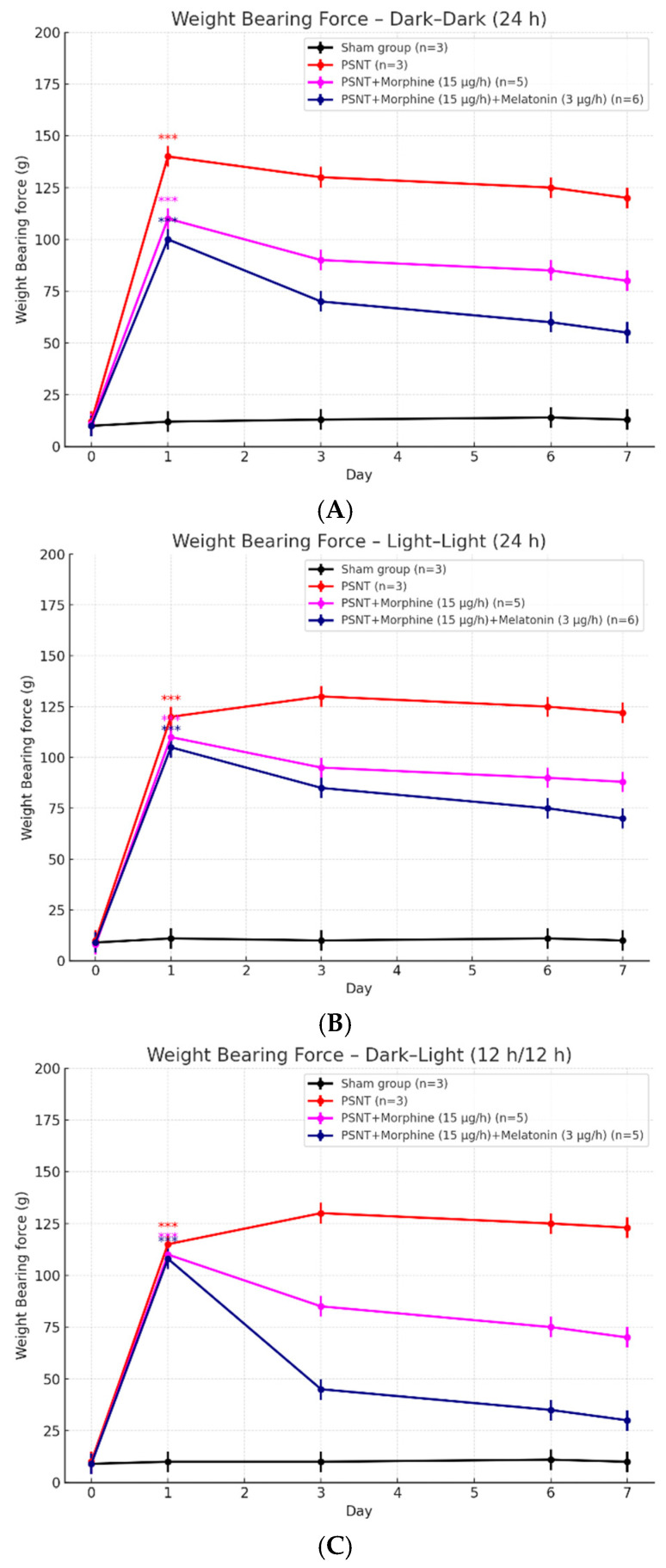
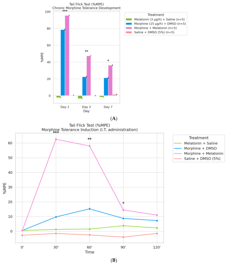
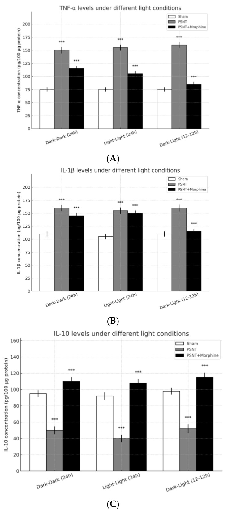
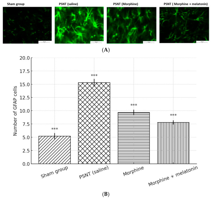
Disruption of circadian rhythms by abnormal light exposure and reduced melatonin secretion has been linked to heightened pain sensitivity and opioid tolerance. This study evaluated how environmental light manipulation and exogenous melatonin supplementation influence pain perception and morphine tolerance in a rat model of neuropathic pain induced by partial sciatic nerve transection (PSNT). Rats were exposed to constant darkness, constant light, or a 12 h/12 h light-dark cycle for one week before PSNT surgery. Behavioral assays and continuous intrathecal (i.t.) infusion of morphine, melatonin, or their combination were conducted over a 7-day period beginning immediately after PSNT. On Day 7, after discontinued drugs infusion, an acute intrathecal morphine challenge (15 µg, i.t.) was administered to assess tolerance expression. Constant light suppressed melatonin levels, exacerbated pain behaviors, and accelerated morphine tolerance. In contrast, circadian-aligned lighting preserved melatonin rhythms and mitigated these effects. Melatonin co-infusion attenuated morphine tolerance and enhanced morphine analgesia. Reduced pro-inflammatory cytokine expression and increase anti-inflammatory cytokine IL-10 level and suppressed astrocyte activation were also observed by melatonin co-infusion during morphine tolerance induction. These findings highlight the potential of melatonin and circadian regulation in improving opioid efficacy and reduced morphine tolerance in managing neuropathic pain.

Keywords: astrocytes; circadian rhythm; light exposure; melatonin; morphine tolerance; neuroinflammation; neuropathic pain; opioid-sparing therapy.


Conflict of interest statement

The authors declare no conflict of interest.

Figures

Figure 1
Figure 1
Circadian variation in serum melatonin levels under different treatment and lighting conditions. Serum melatonin concentrations were measured in rats subjected to various treatments and environmental light exposures. (A) Baseline circadian melatonin rhythm. Serum melatonin levels were assessed over a 12 h dark period (10 p.m. to 10 a.m.) on Day 4, prior to surgery. Three groups were compared: sham, PSNT, and PSNT + morphine (n = 5 per group). All groups exhibited a nocturnal surge in melatonin, peaking between 2–4 a.m. The overall pattern confirmed the presence of an intact circadian rhythm prior to intervention. Data are shown as mean ± SEM. (BD) Melatonin peak levels under different lighting conditions. Post-surgical peak melatonin concentrations (Day 7) under three light exposure paradigms—constant darkness (dark–dark, DD), standard 12 h light–dark cycle (dark–light, DL), and continuous light (light–light, LL)—were compared against baseline values from (A) (light-colored bars). (B) Sham group: Peak melatonin levels were preserved under DD and DL conditions but markedly suppressed under LL exposure (Peak: 11.5 pg/mL), indicating circadian disruption by constant light. (C) PSNT group: Peak melatonin concentrations were moderately reduced across all light conditions, particularly under LL (Peak: 10.7 pg/mL), suggesting nerve injury impairs nocturnal melatonin output. (D) PSNT + morphine group: Melatonin levels were further diminished, especially under LL conditions (Peak: 10.1 pg/mL), indicating a combined suppressive effect of morphine and disrupted circadian cues. Bars represent the mean ± SEM; peak values are annotated in red. *** p < 0.001.
Figure 2
Figure 2
Effects of different light exposure conditions on paw withdrawal threshold for mechanical allodynia measurement in PSNT rats treated with morphine and melatonin. Line plots show the paw withdrawal threshold (g) measured over a 7-day period under three lighting conditions: (A) constant darkness (dark–dark, 24 h), (B) constant light (light–light, 24 h), and (C) standard 12 h light–dark cycle (dark–light, 12 h–12 h). Rats were divided into four groups: sham-operated controls (n = 3), PSNT (partial sciatic nerve transection, n = 3), PSNT + morphine (15 μg/h, n = 6), and PSNT + morphine + melatonin (3 μg/h, n = 6). Data are presented as the mean ± SEM. Statistical comparisons were performed against baseline (Day 0). *** p < 0.001. PSNT significantly reduced withdrawal thresholds under all lighting conditions. Morphine initially reversed this effect, but tolerance developed over time. Co-administration of melatonin preserved analgesic responses, especially under circadian-aligned lighting conditions (dark–dark and dark–light), indicating a modulatory role of melatonin in maintaining opioid efficacy.
Figure 3
Figure 3
Effects of morphine and melatonin on weight-bearing asymmetry in PSNT rats under different lighting conditions. Line graphs illustrate daily changes in hind limb weight-bearing force (g) over a 7-day period following partial sciatic nerve transection (PSNT) in rats exposed to different light environments: (A) constant darkness (dark–dark, 24 h), (B) constant light (light–light, 24 h), and (C) standard 12 h–12 h light–dark cycle (dark–light). Experimental groups included sham controls (n = 3), PSNT (n = 3), PSNT + morphine (15 μg/h, n = 5), and PSNT + morphine + melatonin (3 μg/h, n = 5), with drugs delivered continuously via intrathecal infusion. All graphs show the mean ± SEM. Statistical comparisons were made against the baseline value (Day 0). *** p < 0.001. PSNT induced significant mechanical imbalance, characterized by increased contralateral weight-bearing. Morphine provided transient relief, but analgesic efficacy waned over time, indicating tolerance. Co-infusion of melatonin mitigated this effect and helped maintain weight-bearing symmetry, particularly under circadian-supportive conditions (dark–dark and dark–light).
Figure 4
Figure 4
(A) Chronic morphine administration induces analgesic tolerance, which is attenuated by co-treatment with melatonin. Tail-flick latency was measured on Days 1, 3, and 7 following continuous intrathecal infusion of saline, morphine (15 µg/h), or morphine combined with melatonin (3 µg/h). Data are expressed as %MPE (Maximum Possible Effect), calculated using the formula: %MPE = [(post-drug latency − baseline latency)/(cut-off − baseline latency)] × 100, with a baseline latency of 2 s and a cut-off latency of 10 s. Bars represent the mean ± SEM (n = 5 rats per group). p < 0.001, p < 0.01, p < 0.05 compared to all other treatment groups at the same time point. (B) Melatonin enhances the acute antinociceptive effect of morphine and delays tolerance onset. Tail-flick latency was measured at 0, 30, 60, 90, and 120 min following a single intrathecal dose of morphine (15 µg/h), with or without melatonin (3 µg/h), in PSNT rats. Additional control groups received saline or melatonin alone. Data are presented as %MPE (Maximum Possible Effect), calculated using the formula: %MPE = [(post-drug latency − baseline latency)/(cut-off − baseline latency)] × 100, where baseline latency = 2 s and cut-off = 10 s. Each line represents the mean ± SEM (n = 5 per group). * p < 0.05, ** p < 0.01, and *** p < 0.001, for the morphine + melatonin group compared to all other groups at the corresponding time points (30′, 60′, and 90′).
Figure 5
Figure 5
Effects of light exposure and morphine administration on inflammatory cytokine levels in spinal cord tissue following partial sciatic nerve transection (PSNT). Cytokine concentrations (pg/100 μg protein) were quantified in lumbar spinal cord samples from sham, PSNT, and PSNT + morphine groups under three light conditions: constant darkness (dark–dark, 24 h), constant light (light–light, 24 h), and standard 12 h–12 h light–dark cycle (dark–light). (A) TNF-α levels were significantly elevated in PSNT groups across all lighting conditions and partially reduced by morphine. (B) IL-1β levels mirrored TNF-α changes, with PSNT-induced increases attenuated to varying degrees by morphine treatment. (C) IL-10, an anti-inflammatory cytokine, was suppressed following PSNT but markedly upregulated in the PSNT + morphine group under all lighting conditions. Data are presented as the mean ± SEM (n = 5 per group). *** p < 0.001., relative to the sham group for each lighting condition unless otherwise indicated.
Figure 6
Figure 6
Melatonin attenuates PSNT- and morphine-induced astrocyte activation in the spinal cord. (A) Representative immunofluorescence images of GFAP-positive astrocytes in the dorsal horn of the lumbar spinal cord from four groups: sham, PSNT (saline), PSNT (morphine), and PSNT (morphine + melatonin). Astrocytes are stained green for GFAP. PSNT induced marked astrocytic activation, which was reduced by morphine and further suppressed by co-administration of melatonin. (B) Quantification of GFAP-positive cells per microscopic field. Data are presented as the mean ± SD (n = 5). PSNT significantly increased astrocyte numbers, while morphine and melatonin treatment progressively attenuated this elevation. *** p < 0.001, compared to the PSNT (saline) group unless otherwise indicated.

Similar articles

References

    1. Center for Substance Abuse Treatment . Managing Chronic Pain in Adults with or in Recovery from Substance Use Disorders. Substance Abuse and Mental Health Services Administration (US); Rockville, MD, USA: 2012. SAMHSA/CSAT Treatment Improvement Protocols. - PubMed
    1. Bumgarner J.R., McCray E.W., Nelson R.J. The disruptive relationship among circadian rhythms, pain, and opioids. Front. Neurosci. 2023;17:1109480. doi: 10.3389/fnins.2023.1109480. - DOI - PMC - PubMed
    1. Danilov A., Kurganova J. Melatonin in Chronic Pain Syndromes. Pain Ther. 2016;5:1–17. doi: 10.1007/s40122-016-0049-y. - DOI - PMC - PubMed
    1. Ma H., Yan J., Sun W., Jiang M., Zhang Y. Melatonin Treatment for Sleep Disorders in Parkinson’s Disease: A Meta-Analysis and Systematic Review. Front. Aging Neurosci. 2022;14:784314. doi: 10.3389/fnagi.2022.784314. - DOI - PMC - PubMed
    1. Choy E.H. The role of sleep in pain and fibromyalgia. Nat. Rev. Rheumatol. 2015;11:513–520. doi: 10.1038/nrrheum.2015.56. - DOI - PubMed

LinkOut - more resources