Review
. 2025 Jul 30;26(15):7384.
doi: 10.3390/ijms26157384.

Biomaterial-Based Nucleic Acid Delivery Systems for In Situ Tissue Engineering and Regenerative Medicine

Affiliations
Review

Biomaterial-Based Nucleic Acid Delivery Systems for In Situ Tissue Engineering and Regenerative Medicine

Qi-Xiang Wu et al. Int J Mol Sci. .

Abstract

Gene therapy is a groundbreaking strategy in regenerative medicine, enabling precise cellular behavior modulation for tissue repair. In situ nucleic acid delivery systems aim to directly deliver nucleic acids to target cells or tissues to realize localized genetic reprogramming and avoid issues like donor cell dependency and immune rejection. The key to success relies on biomaterial-engineered delivery platforms that ensure tissue-specific targeting and efficient intracellular transport. Viral vectors and non-viral carriers are strategically modified to enhance nucleic acid stability and cellular uptake, and integrate them into injectable or 3D-printed scaffolds. These scaffolds not only control nucleic acid release but also mimic native extracellular microenvironments to support stem cell recruitment and tissue regeneration. This review explores three key aspects: the mechanisms of gene editing in tissue repair; advancements in viral and non-viral vector engineering; and innovations in biomaterial scaffolds, including stimuli-responsive hydrogels and 3D-printed matrices. We evaluate scaffold fabrication methodologies, nucleic acid loading-release kinetics, and their biological impacts. Despite progress in spatiotemporal gene delivery control, challenges remain in balancing vector biocompatibility, manufacturing scalability, and long-term safety. Future research should focus on multifunctional "smart" scaffolds with CRISPR-based editing tools, multi-stimuli responsiveness, and patient-specific designs. This work systematically integrates the latest methodological advances, outlines actionable strategies for future investigations and advances clinical translation perspectives beyond the existing literature.

Keywords: gene therapy; nanoparticle; nucleic acid delivery system; scaffold; vector.


Conflict of interest statement

The authors declare no conflicts of interest.

Figures
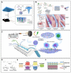
Figure 10
Figure 10
Structure and preparation method of sheet-like scaffolds. (A) The components and application of MN/PBAE/DNA [107]. Copyright 2020, Royal society of chemistry. (B) Fabrication procedure of the siRNA@MS@HA hydrogel–electrospun membrane, and the implantation into rat injured tendon site [223]. Copyright 2022, Wiley-VCH. (C) A schematic representation of a Tesla micromixer to make chitosan-based gene delivery nanocomplex and the electrospinning processes used to make poly(ε-caprolactone) (PCL) nanofibers modified by nanocomplex [86]. Copyright 2020, Elsevier. (D) Chemical structures of Poly2, chitosan and dextran sulfate. (E) The hierarchical structure of LbL films into a single coating. The first (X) film is a hydrolytically degradable undercoating, while the second (Y) film contains the siRNA to be delivered. (F) Architecture of hierarchical LbL [235]. Copyright 2016, Wiley-VCH.
Figure 1
Figure 1
Three mechanisms of gene modification: gene addition, silencing, and CRISPR/Cas9-based genome editing (Created with Cnsknowall.com).
Figure 2
Figure 2
Three key scales of nucleic acid therapeutics for in situ tissue regeneration: tissue targeting, cellular specificity, and intracellular transport.
Figure 3
Figure 3
Schematics of targeting ligands, surface modification, physical properties and materials for nucleic acid delivery vectors. Created with Figdraw (https://www.figdraw.com).
Figure 4
Figure 4
Schematic illustration of the mechanisms of exosomes and lipid-based nucleic acids carriers. (A) Schematic illustration for engineered miR-181b exosomes that improved osteointegration by regulating macrophage polarization [51]. Copyright 2021, BioMed Central. (B) Proposed mechanisms of formation and structure of LNP prepared in the absence and presence of siRNA [57]. Copyright 2018, American Chemical Society. (C) Endosome escape in lipoplex-mediated siRNA delivery [58]. Copyright 2009, Elsevier.
Figure 5
Figure 5
Schematic illustration of the mechanisms of nucleic acid-based nanomaterials. (A) Schematic display of the fabrication of stFNA–miR. (B) Schematic diagram of the enzyme cleavage test in an extracellular environment. (C) Schematic illustration of miRNA transportation into cells by stFNA and selection of a guide strand to realize subsequent biochemical reactions. (D) Flow cytometric results for cellular uptake of stFNA. (E) The binding site of RNase H and the position of the guide and passenger strands [93]. Copyright 2021, Wiley-VCH. (F) The inner core of the BiRDS and the location of the surrounding miRNAs [94]. Copyright 2024, American Chemical Society. (G) The procedure used to prepare miR-5590-SNA@DNAgel [97]. Copyright 2023, BioMed Central.
Figure 6
Figure 6
Designs for stimuli-responsive release and chemical strategies. (A) Synthesis of the photocleavable linker (PL). (B) Chemical strategies for miR-26a conjugation with a photocleavable linker (PL-5) and photosensitivity mechanism [148]. Copyright 2021, Elsevier. (C) Formation of RNA/PEI nanocomplexes and hydrogel fabrication via a single cross-linked Michael addition reaction [181]. Copyright 2017, American Chemical Society. (D) Dual-functional supramolecular hydrogel design for targeted and light-responsive miRNA delivery, including structure transformation between SPI–Gal and MCI–Gal under heat or light control [185]. Copyright 2016, Royal Society of Chemistry. (E) Chemical structure and intra-molecular photo-click reaction of Tet (II) [186]. Copyright 2017, Royal Society of Chemistry. (F) Synthesis of MPEG-PEI-PBLL [187]. Copyright 2020, Royal Society of Chemistry. (G) Formation of miRNA/PGPC polyplex micelles, encapsulation in injectable PEG hydrogels, and molecular mechanism of MMP-2 silencing in nucleus pulposus cells for fibrosis inhibition [150]. Copyright 2018, Wiley-VCH.
Figure 7
Figure 7
Photosensitive and temperature-sensitive hydrogels for biomedical applications. (A) Synthesis of methacrylated gelatin [188]. Copyright 2010, Elsevier. (B) The synthesis and therapeutic effect of the siRNAs/PLNG scaffold for the treatment of SCI [155]. Copyright 2024, American Chemical Society. (C) The structural changes in F127 systems under controlled temperature after flow [190]. Copyright 2020, Elsevier. (D) The synthesis and mechanism of action of the heat-sensitive hydrogel be made up of PLGA–PEG–PLGA [102]. Copyright 2023, AIP publishing. (E) An illustration of how the polymer solution transits from a soluble state to a viscous hydrogel by hydrophobic interactions between the isopropyl groups of pNIPAAM, when the temperature is above its lower critical solution temperature (LCST), 32 °C [191]. Copyright 2015, Elsevier.
Figure 8
Figure 8
Designs of injectable hydrogels for in situ self-assemble and localized release. (A) Chemical structures and cartoon illustration of β-CD and the adamantane derivatives [194]. Copyright 2021, MDPI AG. Licensed under CC BY 4.0. (B) Hyaluronic acid modified by Adamantane and cyclodextrin lead to hydrogel self-assembly via guest–host interactions, and disassembly on shear thinning. MiR-302 modified by cholesterol allows its incorporation into the gel via the guest–host interaction of cholesterol with cyclodextrin [166]. Copyright 2017, Nature Research. (C) Schematic illustration of SKP@miR [167]. Copyright 2022, American Association for the Advancement of Science. Licensed under CC BY-NC 4.0. (D) Fabrication and gelation mechanisms of miR-145-loaded RAD peptide hydrogels [168]. Copyright 2024, Elsevier.
Figure 9
Figure 9
Advances in 3D printing: porous braided scaffolds and bionic structured scaffolds. (A) Schematics illustrating the preparation of a 3D-printed porous braided scaffold loaded with Siv/DPNP/miR-210 and its application for bone regeneration. Reprinted with permission from [129]. Copyright 2021, Royal society of chemistry. (B) Scaffold fabrication schematic diagram. (C,D) SEM imagery illustrating the complete scaffold alongside a magnified view of the PCL electrospun fibers (highlighted in red box). A total of 50 fibers were analyzed, yielding an average diameter of 1.35 ± 0.19 µm. The black arrow, positioned in the top right corner of (C), denotes the orientation of the fibers. (E) Cumulative release of Neg miRNA and GDNF over time [198]. Copyright 2021, Wiley-VCH. Licensed under CC BY 4.0. (F) Schematic representation for preparation of the bilayer electrospun membranes used in vascular tissue engineering [199]. Copyright 2016, Elsevier. (G) Schematic illustration of the composite scaffolds ((INS MPs+VD3) Gel+GAM) [99]. Copyright 2020, Wiley-VCH.

Similar articles

References

    1. Ding S. Therapeutic Reprogramming toward Regenerative Medicine. Chem. Rev. 2025;125:1805–1822. doi: 10.1021/acs.chemrev.4c00332. - DOI - PubMed
    1. Asch A., Kalbermatten D.F., Madduri S. Clinical Safety and Efficacy of Allogeneic Adipose Stem Cells: A Systematic Review of the Clinical Trials. Int. J. Mol. Sci. 2025;26:6376. doi: 10.3390/ijms26136376. - DOI - PMC - PubMed
    1. Chang D., Yang X., Fan S., Fan T., Zhang M., Ono M. Engineering of MSCs Sheet for the Prevention of Myocardial Ischemia and for Left Ventricle Remodeling. Stem Cell Res. Ther. 2023;14:102. doi: 10.1186/s13287-023-03322-7. - DOI - PMC - PubMed
    1. Eweje F., Walsh M.L., Ahmad K., Ibrahim V., Alrefai A., Chen J., Chaikof E.L. Protein-Based Nanoparticles for Therapeutic Nucleic Acid Delivery. Biomaterials. 2024;305:122464. doi: 10.1016/j.biomaterials.2023.122464. - DOI - PMC - PubMed
    1. Cao Q., Fang H., Tian H. mRNA Vaccines Contribute to Innate and Adaptive Immunity to Enhance Immune Response in Vivo. Biomaterials. 2024;310:122628. doi: 10.1016/j.biomaterials.2024.122628. - DOI - PubMed

LinkOut - more resources