. 2025 Jul 30;26(15):7386.
doi: 10.3390/ijms26157386.

Accumulation Kinetics and Biological Action of Doxorubicin in Rabbit Intervertebral Discs

Affiliations

Accumulation Kinetics and Biological Action of Doxorubicin in Rabbit Intervertebral Discs

Eleni Mavrogonatou et al. Int J Mol Sci. .

Abstract

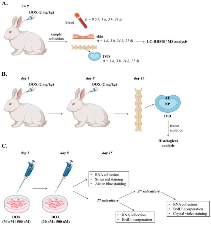
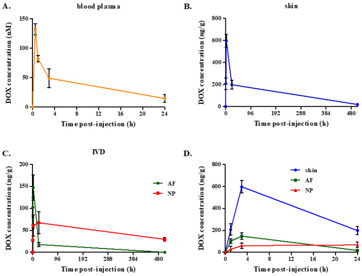
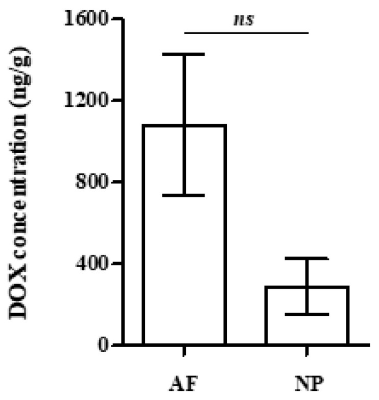
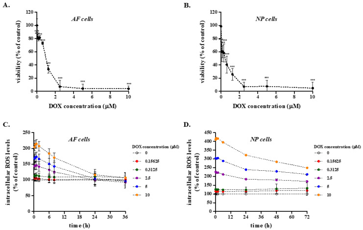
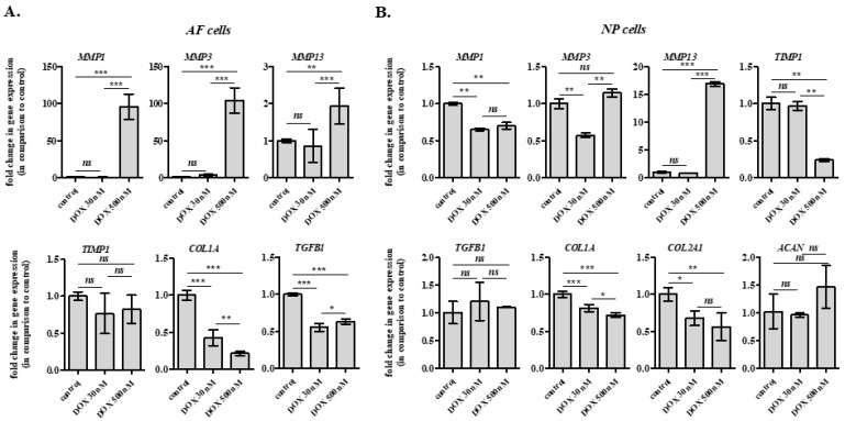
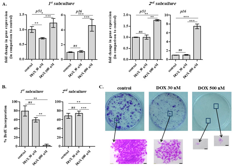
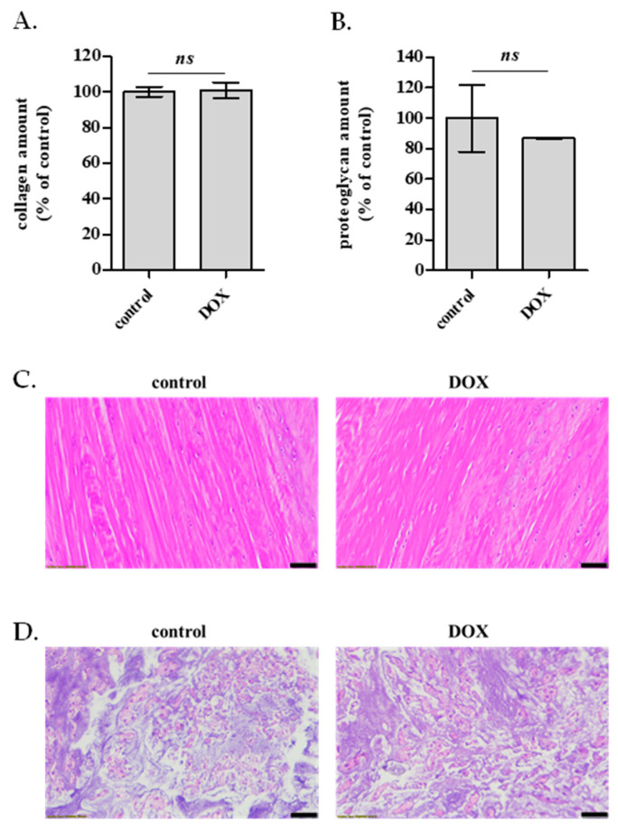
Doxorubicin (DOX) is widely used for the treatment of several tumors, but considerable dose-dependent side effects on many normal tissues, including bones, have been reported. The aim of the present study was to follow for the first time the kinetics of DOX accumulation/clearance in the non-vascularized intervertebral disc (IVD), as well as to assess the drug's biological action in the annulus fibrosus (AF) and nucleus pulposus (NP) IVD cells and tissues. DOX was administered intravenously to rabbits before the isolation of IVDs, in which DOX quantification was performed using a highly sensitive LC-HRMS/MS analytical method. The effect of the drug on IVD cells' physiology was assessed in vitro, while IVD tissue quality post-DOX administration was studied in vivo through histological analysis. DOX delivery was found significantly lower in the IVD compared to the highly vascularized skin, declining from the outer AF to the inner NP. The low DOX concentrations reaching the IVDs had marginal effects on cells' viability, intracellular redox status, and p38 MAPK activation, while they did not evoke cellular senescence. Most importantly, the drug did not negatively affect ECM integrity, as collagen and proteoglycan content remained stable in vitro and in vivo.

Keywords: annulus fibrosus; cell viability; collagen; in vitro; in vivo; nucleus pulposus; oxidative stress; proteoglycans; senescence.


Conflict of interest statement

The authors declare no conflicts of interest. The funders had no role in the design of the study; in the collection, analyses, or interpretation of data; in the writing of the manuscript; or in the decision to publish the results.

Figures

Figure 1
Figure 1
Chemical structure of doxorubicin (DOX).
Figure 2
Figure 2
Graphical representation of doxorubicin administration protocols employed in in vivo and in vitro experiments of the current study. (A). Doxorubicin (DOX) was administered to rabbits intravenously through the ear vein at a dose of 2 mg/kg before blood plasma, skin and intervertebral disc (IVD) (annulus fibrosus, AF and nucleus pulposus, NP) isolation at the designated time-points, processing and preparation for LC-HRMS/MS analysis. (B). DOX was administered to the animals intravenously in two doses of 2 mg/kg on days 1 and 8. Untreated animals served as the control group. DOX-treated and control animals were euthanized at day 15 for tissue isolation and histological analysis. (C). AF and NP cell cultures were treated with 30 and 500 nM of DOX twice, with a 7-day interval, before gene expression analysis and assessment of collagen amount and proteoglycans’ content 7 days post-second DOX administration. For the assessment of DOX-induced cellular senescence, cells were subcultured once and twice (starting 7 days post-second DOX administration) before gene expression and cell proliferation analyses.
Figure 3
Figure 3
Kinetics of doxorubicin (DOX) accumulation in the blood plasma, skin and intervertebral disc (IVD) (annulus fibrosus, AF and nucleus pulposus, NP). DOX was administered to rabbits intravenously at a dose of 2 mg/kg before sample collection at the designated time-points, processing and liquid chromatography-high resolution mass spectrometry (LC-HRMS)/MS analysis. (A). Plasma was isolated by venous blood samples obtained from the ear that did not receive DOX. (B,C). Hairless skin and IVD tissue samples were isolated from DOX-treated and control animals. IVDs were separated from the bilateral vertebral bodies before NP and AF segregation. Tissue samples were collagenase-digested, centrifuged and supernatants were processed for LC-HRMS/MS analysis. (D). Merged representation of (B,C) at the time frame of 0–24 h post-injection, for the simultaneous monitoring/comparison of DOX accumulation in the vascularized skin and avascular IVD tissues. Averages ± standard deviations of three experiments are presented here.
Figure 4
Figure 4
Uptake of doxorubicin (DOX) by annulus fibrosus (AF) and nucleus pulposus (NP) intervertebral disc (IVD) tissues immersed in a solution of DOX at the concentration of the rabbit plasma. In order to assess the uptake of DOX in tissues, AF and NP IVD samples post-immersion were treated with 2 mL of collagenase solution (3 mg/mL). The samples were left overnight under continuous shaking, were then centrifuged, and the supernatant was used for solid phase extraction (SPE) treatment and liquid chromatography-high resolution mass spectrometry (LC-HRMS)/MS analysis. No statistically significant differences were found between samples (Student’s t-test, ns: not significant).
Figure 5
Figure 5
Effect of doxorubicin (DOX) on the viability and redox status of primary rabbit annulus fibrosus (AF) and nucleus pulposus (NP) intervertebral disc (IVD) cells. (A,B). Rabbit AF and NP IVD cells were plated in 96-well plates before the addition of DOX at concentrations from 0 to 10 μΜ for 72 h. Optical density of the solubilized formed MTT formazan crystals was measured at 550 nm and cell viability was calculated as a percent ratio of untreated cells. One representative experiment conducted in sextuplicate is depicted here, and the data presented are mean values ± standard deviations. Asterisks denote statistically significant differences compared to the untreated control (Student’s t-test, *** p < 0.001). (C,D). Rabbit AF and NP IVD cells were cultured in 96-well plates and incubated with 10 μM DCFH-DA for 1 h at 37 °C before their exposure to DOX (0–10 μΜ). Intracellular levels of reactive oxygen species (ROS) were estimated by recording fluorescence (excitation wavelength: 485 nm, emission wavelength: 520 nm). ROS production was expressed as a % ratio of the untreated control. Experiment was repeated three independent times, and one representative graph is shown here.
Figure 6
Figure 6
Doxorubicin (DOX) concentration prevailing in the plasma slightly affects the transcriptional profile of rabbit annulus fibrosus (AF) and nucleus pulposus (NP) intervertebral disc (IVD) cells in vitro. AF (A) and NP (B) cells were treated twice with 30 or 500 nM DOX with a 7-day interval before sample collection and RNA extraction 7 days post-second DOX administration. Extracted RNA was then used for RT-qPCR analysis, with glyceraldehyde-3-phosphate dehydrogenase (GAPDH) serving as the reference gene. Numerical values are the means ± standard deviations of at least two independent experiments conducted in duplicate. Asterisks denote statistically significant differences between any pair of means (ANOVA, Tukey’s test, * p < 0.05, ** p < 0.01, *** p < 0.001, ns: not significant).
Figure 7
Figure 7
Doxorubicin (DOX) concentration prevailing in the plasma does not result in the induction of IVD cells’ senescence in vitro. DOX 30 and 500 nM were administered to rabbit annulus fibrosus (AF) intervertebral disc (IVD) cells once per week for two weeks, and 7 days post-second DOX addition, cells were subcultured once or twice. (A). RNA was extracted 7 days post-subculture and RT-qPCR analysis for p53 and p16INK4a (p16) was performed, with glyceraldehyde-3-phosphate dehydrogenase (GAPDH) serving as the reference gene. (B). Cells exposed to DOX treatment following the same protocol were plated onto coverslips before their incubation with bromodeoxyuridine (BrdU), fixation and immunofluorescence analysis for the estimation of BrdU incorporation. Numerical values are the means ± standard deviations of at least two independent experiments conducted in duplicate. Asterisks denote statistically significant differences between any pair (ANOVA, Tukey’s test, * p < 0.05, ** p < 0.01, *** p < 0.001, ns: not significant). (C). Rabbit AF IVD cells after the second subculture were sparsely plated in 6-well plates. Cells were stained with Crystal violet two weeks later. Scale bar: 200 μm.
Figure 8
Figure 8
Effect of doxorubicin (DOX) on rabbit annulus fibrosus (AF) and nucleus pulposus (NP) extracellular matrix (ECM) quality in vitro and in vivo. (A). Collagen deposition in rabbit AF cells cultured in the absence (control) or the presence of DOX (30 nM) administered twice with a 7-day interval was assessed with Sirius Red staining 7 days post-second DOX administration. Quantification of solubilized collagen-bound Sirius Red stain was performed by measuring the absorbance at 540 nm. (B). Glycosaminoglycans’ content in rabbit NP IVD cells cultured in the absence (control) or the presence of DOX (30 nM) administered twice with a 7-day interval was assessed with Alcian Blue staining 7 days post-second DOX administration. Quantification of the extracted Alcian Blue stain was performed by measuring the absorbance at 620 nm. Collagen amount and glycosaminoglycans’ content normalized to cell number were expressed as a % ratio of the control. Mean values ± standard deviations of a representative experiment conducted in triplicate are demonstrated. No statistically significant differences were observed compared to the untreated control (Student’s t-test, ns: not significant). (C,D). Five μm histological sections of formalin-fixed and paraffin-embedded rabbit AF and NP IVD tissues, derived from the lumbar spine of animals injected twice with 2 mg/kg DOX 7 days post-second DOX injection, were mounted on microscopic slides and stained with hematoxylin or eosin. AF (C) and NP (D) IVD sections were examined under transmitted light microscopy. Representative digital photos obtained using ×20 objective lenses are shown here. Scale bar: 50 μm.

Similar articles

References

    1. Yun U.J., Lee J.H., Shim J., Yoon K., Goh S.H., Yi E.H., Ye S.K., Lee J.S., Lee H., Park J., et al. Anti-cancer effect of doxorubicin is mediated by downregulation of HMG-Co A reductase via inhibition of EGFR/Src pathway. Lab. Investig. 2019;99:1157–1172. doi: 10.1038/s41374-019-0193-1. - DOI - PubMed
    1. Doroshow J.H. Doxorubicin-induced cardiac toxicity. N. Engl. J. Med. 1991;324:843–845. doi: 10.1056/NEJM199103213241210. - DOI - PubMed
    1. Rana T., Chakrabarti A., Freeman M., Biswas S. Doxorubicin-mediated bone loss in breast cancer bone metastases is driven by an interplay between oxidative stress and induction of TGFβ. PLoS ONE. 2013;8:e78043. doi: 10.1371/annotation/95cefb34-2f3d-42a5-b73e-53c531591f0b. - DOI - PMC - PubMed
    1. Shusterman S., Meadows A.T. Long term survivors of childhood leukemia. Curr. Opin. Hematol. 2000;7:217–222. doi: 10.1097/00062752-200007000-00004. - DOI - PubMed
    1. Hadji P., Ziller M., Maskow C., Albert U., Kalder M. The influence of chemotherapy on bone mineral density, quantitative ultrasonometry and bone turnover in pre-menopausal women with breast cancer. Eur. J. Cancer. 2009;45:3205–3212. doi: 10.1016/j.ejca.2009.09.026. - DOI - PubMed

Substances

LinkOut - more resources