Review
. 2025 Jul 31;26(15):7393.
doi: 10.3390/ijms26157393.

Muscarinic Receptor Antagonism and TRPM3 Activation as Stimulators of Mitochondrial Function and Axonal Repair in Diabetic Sensorimotor Polyneuropathy

Affiliations
Review

Muscarinic Receptor Antagonism and TRPM3 Activation as Stimulators of Mitochondrial Function and Axonal Repair in Diabetic Sensorimotor Polyneuropathy

Sanjana Chauhan et al. Int J Mol Sci. .

Abstract

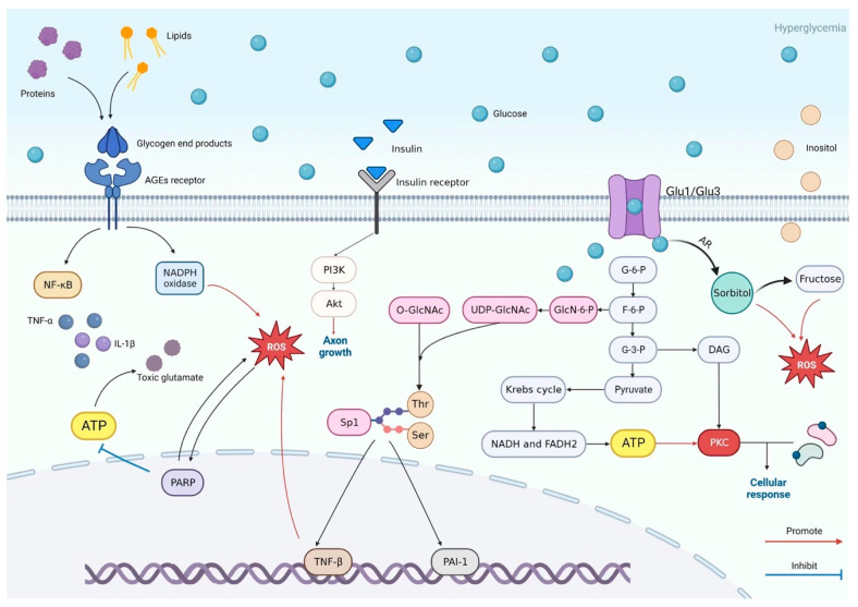
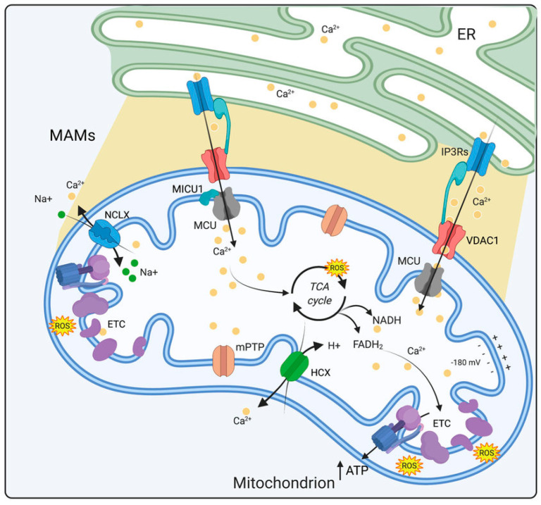
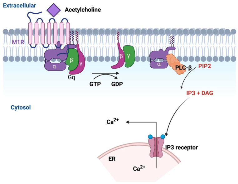
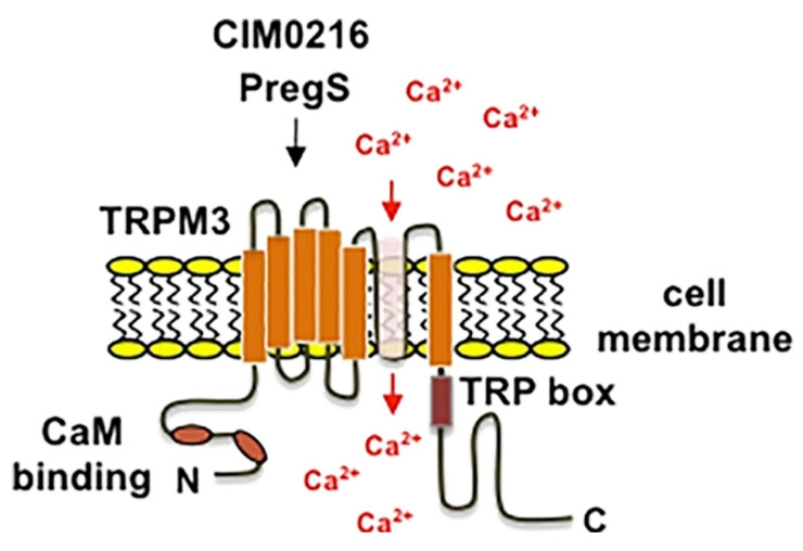
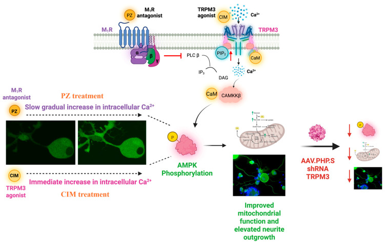
Diabetic sensorimotor polyneuropathy (DSPN) is the most prevalent complication of diabetes, affecting nearly half of all persons with diabetes. It is characterized by nerve degeneration, progressive sensory loss and pain, with increased risk of ulceration and amputation. Despite its high prevalence, disease-modifying treatments for DSPN do not exist. Mitochondrial dysfunction and Ca2+ dyshomeostasis are key contributors to the pathophysiology of DSPN, disrupting neuronal energy homeostasis and initiating axonal degeneration. Recent findings have demonstrated that antagonism of the muscarinic acetylcholine type 1 receptor (M1R) promotes restoration of mitochondrial function and axon repair in various neuropathies, including DSPN, chemotherapy-induced peripheral neuropathy (CIPN) and HIV-associated neuropathy. Pirenzepine, a selective M1R antagonist with a well-established safety profile, is currently under clinical investigation for its potential to reverse neuropathy. The transient receptor potential melastatin-3 (TRPM3) channel, a Ca2+-permeable ion channel, has recently emerged as a downstream effector of G protein-coupled receptor (GPCR) pathways, including M1R. TRPM3 activation enhanced mitochondrial Ca2+ uptake and bioenergetics, promoting axonal sprouting. This review highlights mitochondrial and Ca2+ signaling imbalances in DSPN and presents M1R antagonism and TRPM3 activation as promising neuro-regenerative strategies that shift treatment from symptom control to nerve restoration in diabetic and other peripheral neuropathies.

Keywords: Ca2+ homeostasis; DRG; GPCR; bioenergetics; diabetic neuropathy; pirenzepine.


Conflict of interest statement

The corresponding author, P.F., and co-author N.A.C. declare that they are co-founders of, and shareholders in, WinSanTor Inc., a biotechnology company which has licensed intellectual property from the University of Manitoba and UCSD in the area of antimuscarinic drugs.

Figures

Figure 1
Figure 1
Hyperglycemia-driven pathways contributing to DSPN. Under hyperglycemic conditions, excess glucose passively enters cells through glucose transporters and is shunted into multiple metabolic pathways. Glucose is converted into sorbitol and fructose via the polyol pathway, generating reactive oxygen species (ROS) and redox imbalance. Increased glucose also promotes the formation of advanced glycation end-products (AGEs), activating their receptor and stimulating NADPH oxidase, NF-κB, and pro-inflammatory cytokines. The hexosamine pathway modifies transcription factors (e.g., Sp1), altering gene expression of factors such as TNF-β. Elevated diacylglycerol (DAG) activates protein kinase C (PKC), influencing cellular responses. Collectively, these intertwined mechanisms lead to oxidative stress, inflammation, and disrupted cellular function. Reprinted/Adapted from ref. [34].
Figure 2
Figure 2
Mitochondrial Ca2+ homeostasis. Mitochondrial Ca2+ homeostasis is tightly regulated by influx and efflux mechanisms. Ca2+ enters the mitochondrial matrix via the MCU and through a high electronegative potential (−180 mV) while its extrusion depends on NCLX and HCX exchangers. Within the matrix, Ca2+ stimulates the activity of three dehydrogenases of the Krebs cycle and ATP production. Ca2+ ions are depicted as yellow dots. Abbreviations: ER, endoplasmic reticulum; MAMs, mitochondria associated membranes; ETC, electron transport chain; MCU, mitochondrial Ca2+ uniporter; VDAC1, voltage-dependent anion channel 1; ATP, adenosine triphosphate; MICU1, mitochondrial Ca2+ uptake 1; IP3Rs, inositol-1,4,5-trisphosphate receptors; ROS, reactive oxygen species; mPTP, mitochondrial permeability transition pore; NCLX, Na+/Ca2+ exchanger; HCX, H+/Ca2+ exchanger. Reprinted/Adapted from ref. [46].
Figure 3
Figure 3
AMPK domains and structure. (a) Domain organization of AMPK subunits. Residue numbering refers to human α1, β1 and γ1 isoforms. The α subunit consists of an N-terminal kinase domain, an autoinhibitory sequence [83] and a β-subunit interacting domain (β-SID). The β subunit is N-terminally myristoylated (myr) and contains a mid-molecule carbohydrate-binding module (CBM) and C-terminal αγ subunit-binding sequence (SBS). The γ subunit contains four cystathione β-synthase (CBS) domains, paired (1+2 and 3+4) to form two Bateman modules. (b) Tetrad organization of CBS domains in the γ-subunit, colored as in (a), showing locations of nucleotide binding sites (black arrows). (c) Structure of the mammalian AMPK regulatory core and kinase domain [PDB 2Y94: rat α1 (7–299)/(331–469)/(524–548), human β1 (198–272) (green), rat γ1 (23–326) (red)]; α-subunit regions are colored as in (a). AMP bound at γ site 3 is evident. Reprinted/Adapted with permission from ref. [84]. 2012 Jonathan S. et al.
Figure 4
Figure 4
Schematic of M1 muscarinic receptor (M1R) signaling via the Gq protein pathway. When acetylcholine (ACh) binds to M1R, the Gq heterotrimeric protein (α, β, γ) becomes activated, exchanging GDP for GTP on the α-subunit. The activated Gqα then stimulates phospholipase Cβ (PLCβ), which cleaves the membrane lipid phosphatidylinositol 4,5-bisphosphate (PIP2) into two second messengers: inositol 1,4,5-trisphosphate (IP3) and diacylglycerol (DAG). IP3 diffuses through the cytosol and binds to IP3 receptors on the endoplasmic reticulum (ER), triggering Ca2+ ion (Ca2+) release into the cytosol. Increased cytosolic Ca2+, along with DAG, mediate downstream signaling events that lead to various cellular responses. Created with BioRender. Sanjana Chauhan. (2025) https://BioRender.com/akv9wt9 (accessed on 28 May 2025).
Figure 5
Figure 5
Modular structure, membrane topology, and expression of TRPM3. TRPM3 has six transmembrane domains with a pore-forming domain between transmembrane regions 5 and 6. Both the N- and C-termini project into the cytosol. The N-terminus contains two calmodulin binding sites encompassing amino acids 35–124 and 291–382. The C-terminus contains the TRP domain on the C-terminal side of the sixth transmembrane domain. Reprinted/Adapted with permission from ref. [148]. 2017 Gerald Thiel et al.
Figure 6
Figure 6
Schematic representation of TRPM3 activation and its role in M1R antagonist-mediated neuroprotection. M1R antagonism (PZ) prevents PIP2 hydrolysis, leading to a gradual increase in intracellular Ca2+, whereas TRPM3 agonists (CIM0216/PS) induce an immediate Ca2+ influx. Increased Ca²⁺ activates CaMKKβ-mediated AMPK phosphorylation, enhancing mitochondrial function and neurite outgrowth. Dashed lines indicate connections between representative cellular images and their respective Ca2+ signaling outcomes. Right panel shows that TRPM3 knockdown reduces AMPK activation and mitochondrial bioenergetics and metabolism. Created with BioRender.Sanjana Chauhan. (2025) https://BioRender.com/jkmai3w (accessed on 28 May 2025).

Similar articles

References

    1. Eid S.A., Rumora A.E., Beirowski B., Bennett D.L., Hur J., Savelieff M.G., Feldman E.L. New perspectives in diabetic neuropathy. Neuron. 2023;111:2623–2641. doi: 10.1016/j.neuron.2023.05.003. - DOI - PMC - PubMed
    1. Sloan G., Selvarajah D., Tesfaye S. Pathogenesis, diagnosis and clinical management of diabetic sensorimotor peripheral neuropathy. Nat. Rev. Endocrinol. 2021;17:400–420. doi: 10.1038/s41574-021-00496-z. - DOI - PubMed
    1. Rumora A.E., Kim B., Feldman E.L. A Role for Fatty Acids in Peripheral Neuropathy Associated with Type 2 Diabetes and Prediabetes. Antioxid. Redox Signal. 2022;37:560–577. doi: 10.1089/ars.2021.0155. - DOI - PMC - PubMed
    1. Zochodne D.W. Sensory Neurodegeneration in Diabetes: Beyond Glucotoxicity. Int. Rev. Neurobiol. 2016;127:151–180. doi: 10.1016/bs.irn.2016.03.007. - DOI - PubMed
    1. Miyashita A., Kobayashi M., Yokota T., Zochodne D.W. Diabetic Polyneuropathy: New Strategies to Target Sensory Neurons in Dorsal Root Ganglia. Int. J. Mol. Sci. 2023;24:5977. doi: 10.3390/ijms24065977. - DOI - PMC - PubMed

LinkOut - more resources