. 2025 Jul 31;26(15):7404.
doi: 10.3390/ijms26157404.

Effects of Near-Infrared Diode Laser Irradiation on Pain Relief and Neuropeptide Markers During Experimental Tooth Movement in the Periodontal Ligament Tissues of Rats: A Pilot Study

Affiliations

Effects of Near-Infrared Diode Laser Irradiation on Pain Relief and Neuropeptide Markers During Experimental Tooth Movement in the Periodontal Ligament Tissues of Rats: A Pilot Study

Kanako Okazaki et al. Int J Mol Sci. .

Abstract

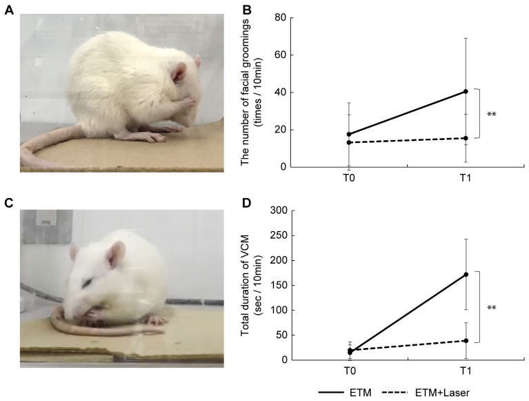
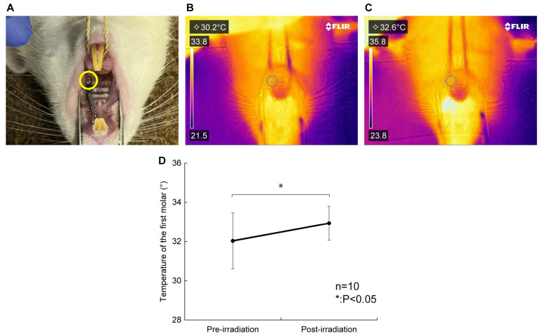
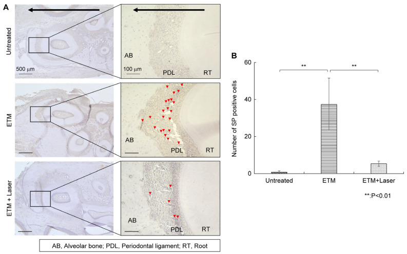
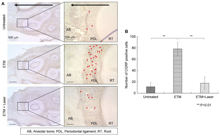
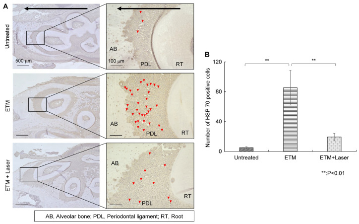
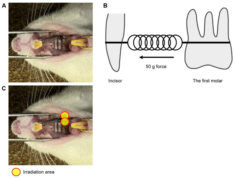
Pain following orthodontic treatment is the chief complaint of patients undergoing this form of treatment. Although the use of diode lasers has been suggested for pain reduction, the mechanism of laser-induced analgesic effects remains unclear. Neuropeptides, such as substance P (SP) and calcitonin gene-related peptide (CGRP), contribute to the transmission and maintenance of inflammatory pain. Heat shock protein (HSP) 70 plays a protective role against various stresses, including orthodontic forces. This study aimed to examine the effects of diode laser irradiation on neuropeptides and HSP 70 expression in periodontal tissues induced by experimental tooth movement (ETM). For inducing ETM for 24 h, 50 g of orthodontic force was applied using a nickel-titanium closed-coil spring to the upper left first molar and the incisors of 20 male Sprague Dawley rats (7 weeks old). The right side without ETM treatment was considered the untreated control group. In 10 rats, diode laser irradiation was performed on the buccal and palatal sides of the first molar for 90 s with a total energy of 100.8 J/cm2. A near-infrared (NIR) laser with a 808 nm wavelength, 7 W peak power, 560 W average power, and 20 ms pulse width was used for the experiment. We measured the number of facial groomings and vacuous chewing movements (VCMs) in the ETM and ETM + laser groups. Immunohistochemical staining of the periodontal tissue with SP, CGRP, and HSP 70 was performed. The number of facial grooming and VCM periods significantly decreased in the ETM + laser group compared to the ETM group. Moreover, the ETM + laser group demonstrated significant suppression of SP, CGRP, and HSP 70 expression. These results suggest that the diode laser demonstrated analgesic effects on ETM-induced pain by inhibiting SP and CGRP expression, and decreased HSP 70 expression shows alleviation of cell damage. Thus, although further validation is warranted for human applications, an NIR diode laser can be used for reducing pain and neuropeptide markers during orthodontic tooth movement.

Keywords: calcitonin gene-related peptide; heat shock protein 70; near-infrared diode laser; neuropeptides; periodontal tissues; photobiomodulation; substance P.


Conflict of interest statement

The authors declare no conflicts of interest.

Figures

Figure 1
Figure 1
Effects of diode laser irradiation on nociceptor behavior (facial grooming and vacuous chewing movement) during experimental tooth movement. (A) Images captured from a video showing the facial grooming of rats in pain. (B) The number of facial grooming events in the experimental tooth movement (ETM) + laser group at T1 was significantly lower than that in the ETM group at T1 (p < 0.01). At T0, no significant differences were observed between the ETM and ETM + laser groups (p = 0.499). (C) Images captured from a video showing the vacuous chewing movement of the rats in pain. (D) The vacuous chewing movement period in the ETM + laser group at T1 was significantly shorter than that in the ETM group at T1 (p < 0.01). At T0, no significant differences were observed between the ETM and ETM + laser groups (p = 0.718). n = 10. ** p < 0.01 (Tukey’s test).
Figure 2
Figure 2
Effects of diode laser irradiation on temperature. (A) The temperature of the upper left first molar of the rat, the irradiated site (yellow circle), was measured. (B) FLIR camera thermogram before irradiation. The measured temperatures are shown in the upper left. (C) FLIR camera thermogram after irradiation. The measured temperatures are shown in the upper left. (D) The temperature of the periodontal tissue increased significantly post-irradiation compared with pre-irradiation (32.0 ± 1.4 vs. 32.9 ± 0.9 °C, p < 0.05). The average temperature change was +0.9 °C, and no increase of more than 2.4 °C was observed. n = 10. * p < 0.05, compared to pre-irradiation (Wilcoxon signed-rank test).
Figure 3
Figure 3
Immunohistochemical staining for substance P. Expression of substance P in the periodontal tissues. (A) As tooth movement occurs in the direction of the arrow (black) in the figure, the left side of each root was considered the compression side. The arrowheads indicate P-positive cells (red). (B) The number of substance P-positive cells in the periodontal ligament on the compression side in the untreated, ETM, and ETM + laser groups. n = 5. ** p < 0.01 (Tukey’s tests). Scale bar, 500 μm in the left-hand slides (×4); 100 μm in the right-hand slides (×20). AB, alveolar bone; PDL, periodontal ligament; RT, root.
Figure 4
Figure 4
Immunohistochemical staining. CGRP expression in the periodontal tissues. (A) As tooth movement occurs in the direction of the arrow (black) in the figure, the left side of each root was considered the compression side. The arrowheads indicate CGRP-positive cells (red). (B) The number of CGRP-positive cells in the periodontal ligament on the compression side in the untreated, ETM, and ETM + laser groups. n = 5. ** p < 0.01 (Tukey’s tests). Scale bar, 500 μm in the left-hand slides (×4); 100 μm in the right-hand slides (×20). CGRP, calcitonin gene-related peptide; AB, alveolar bone; PDL, periodontal ligament; RT, root.
Figure 5
Figure 5
HSP 70 immunohistochemical staining. The expression of HSP 70 in the periodontal tissues. (A) As tooth movement occurs in the direction of the arrow (black) in the figure, the left side of each root was considered the compression side. Arrowheads indicate HSP 70-positive cells (red). (B) Number of HSP 70-positive cells in the periodontal ligament on the compression side in the untreated, ETM, and ETM + laser groups. n = 5. ** p < 0.01 (Tukey’s tests). Scale bar, 500 μm in the left-hand slides (×4); 100 μm in the right-hand slides (×20). HSP 70, heat shock protein 70; AB, alveolar bone; PDL, periodontal ligament; RT, root.
Figure 6
Figure 6
Experimental tooth model. (A) A 50-gF nickel–titanium (Ni-Ti) closed-coil spring was applied between the upper left first molar and upper incisors of the rat using an orthodontic ligature wire. (B) The buccal and palatal sides of the first molar were irradiated for 45 s in each area, and the total energy was set to 100.8 J/cm2. (C) In vivo model of experimental tooth movement. A Ni-Ti closed-coil spring was used, and the orthodontic force was set to 50 g. The ligatures were bonded with resin cement to prevent detachment of the appliance.

Similar articles

References

    1. Long H., Wang Y., Jian F., Liao L.-N., Yang X., Lai W.-L. Current advances in orthodontic pain. Int. J. Oral Sci. 2016;8:67–75. doi: 10.1038/ijos.2016.24. - DOI - PMC - PubMed
    1. Krishnan V. Orthodontic pain: From causes to management—a review. Eur. J. Orthod. 2007;29:170–179. doi: 10.1093/ejo/cjl081. - DOI - PubMed
    1. Kavaliauskiene A., Smailiene D., Buskiene I., Keriene D. Pain and discomfort perception among patients undergoing orthodontic treatment: Results from one month follow-up study. Stomatologija. 2012;14:118–125. - PubMed
    1. Krukemeyer A.M., Arruda A.O., Rohr Inglehart M. Pain and Orthodontic Treatment Patient Experiences and Provider Assessments. Angle Orthod. 2009;79:1175–1181. doi: 10.2319/121308-632R.1. - DOI - PubMed
    1. Wang S., Ko C.C., Chung M.K. Nociceptor mechanisms underlying pain and bone remodeling via orthodontic forces: Toward no pain, big gain. Front. Pain Res. 2024;5:1365194. doi: 10.3389/fpain.2024.1365194. - DOI - PMC - PubMed

MeSH terms

LinkOut - more resources