. 2025 Jul 31;26(15):7406.
doi: 10.3390/ijms26157406.

A Conserved N-Terminal Di-Arginine Motif Stabilizes Plant DGAT1 and Modulates Lipid Droplet Organization

Affiliations

A Conserved N-Terminal Di-Arginine Motif Stabilizes Plant DGAT1 and Modulates Lipid Droplet Organization

Somrutai Winichayakul et al. Int J Mol Sci. .

Abstract

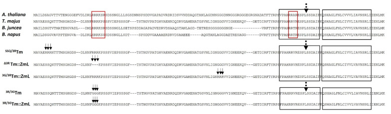
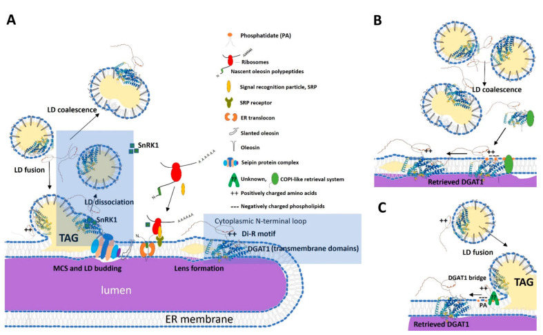
Diacylglycerol-O-acyltransferase 1 (DGAT1, EC 2.3.1.20) is a pivotal enzyme in plant triacylglycerol (TAG) biosynthesis. Previous work identified conserved di-arginine (R) motifs (R-R, R-X-R, and R-X-X-R) in its N-terminal cytoplasmic acyl-CoA binding domain. To elucidate their functional significance, we engineered R-rich sequences in the N-termini of Tropaeolum majus and Zea mays DGAT1s. Comparative analysis with their respective non-mutant constructs showed that deleting or substituting R with glycine in the N-terminal region of DGAT1 markedly reduced lipid accumulation in both Camelina sativa seeds and Saccharomyces cerevisiae cells. Immunofluorescence imaging revealed co-localization of non-mutant and R-substituted DGAT1 with lipid droplets (LDs). However, disruption of an N-terminal di-R motif destabilizes DGAT1, alters LD organization, and impairs recombinant oleosin retention on LDs. Further evidence suggests that the di-R motif mediates DGAT1 retrieval from LDs to the endoplasmic reticulum (ER), implicating its role in dynamic LD-ER protein trafficking. These findings establish the conserved di-R motifs as important regulators of DGAT1 function and LD dynamics, offering insights for the engineering of oil content in diverse biological systems.

Keywords: Camelina; DGAT1; di-arginine motif; lipid droplets; oleosin; transmembrane protein; yeast.


Conflict of interest statement

All authors declare that the research was conducted in the absence of any commercial or financial relationships that could be construed as a potential conflict of interest.

Figures

Figure 1
Figure 1
The alignment shows partial N-terminal cytoplasmic sequences (121 residues) of DGAT1 from Brassicales species, including A. thaliana (total protein: 522 residues, 9 TMDs), T. majus (total protein: 520 residues, 9 TMDs), B. juncea, and B. napus. Red boxes indicate the conserved di-arginine motif (R-R, R-X-R or R-X-X-R-) near the N-terminus and a second motif (RR or R-R-X-R) in the acyl Co-A binding region. The left-hand black box represents the acyl-CoA binding domain [29], while the right-hand box represents the predicted first transmembrane domain [30]. The dashed arrow indicates the engineered site utilized to generate chimeric Tm::ZmL DGAT1 [35]. The solid arrows indicate the site-specific mutation. For expression in C. sativa, the following mutants were used: SSQ/3RTm, representing the substituted S6R, S7R, and S8R mutant of Tm; ∆3RTm::ZmL, representing the deleted R25, R26, and R27 mutant of Tm::ZmL; 3G/3RTm::ZmL, representing the substituted G78R, G79R, and G80R mutant of Tm::ZmL. For expression in S. cerevisiae, the following mutants were used: 3R/3GTm and 3R/3GTm::ZmL, which represent the substituted R25G, R26G, and R27G mutant of Tm and Tm::ZmL, respectively. Accession numbers for the mentioned species are as follows: A. thaliana, NP_179535; T. majus, AAM03340; B. juncea, AAY40785; B. napus, AAD45536; Tm::ZmL, ON959593.
Figure 2
Figure 2
Immunoblot analysis of recombinant DGAT1s in Saccharomyces cerevisiae. The figure displays an immunoblot analysis conducted on microsomal proteins extracted from S. cerevisiae cultures at different time points: 8, 24, and 48 h. Gel loading was meticulously controlled based on equal amounts of total microsomal protein, as demonstrated by the Tris-Glycine eXtended (TGX) stain-free gel in the top panel. Quantities of the Kar2 reference protein [44] are indicated by the ~75 kDa band at the bottom membrane of each immunoblot. Recombinant DGAT1 monomers are denoted by single black asterisks. The expected monomeric sizes (in kDa) are as follows: (A) Tm::ZmL (62.8) and 3R/3GTm::ZmL (62.5); (B) Tm (62.6) and 3R/3GTm (62.3). Dimerized DGAT1s are marked with double black asterisks. VC stands for vector control. VC+, Tm::ZmL+, and 3R/3GTm::ZmL+ signify co-expression of DGAT1 with recombinant Camelina sativa oleosin. It is worth noting that the recombinant DGAT1s migrated 15–20% faster in SDS-PAGE than their predicted molecular weights. Additionally, the substitution of R25G, R26G, and R27G resulted in a slightly smaller protein size of 0.3 kDa. However, both 3R/3GTm::ZmL and 3R/3GTm migrated slower than their corresponding native forms. This phenomenon, referred to as “gel shifting” by Rath et al. (2009), is common for membrane proteins due to their non-polar/polar nature [45,46].
Figure 3
Figure 3
Influence of expressing Tropaeolum majus DGAT1 (Tm), chimeric T. majus and Zea mays long-form DGAT1s (Tm::ZmL), and di-arginine mutated DGAT1s (3R/3GTm::ZmL, and 3R/3GTm) in Saccharomyces cerevisiae quadruple mutant strain H1246 [41] on fatty acid (FA) content (A,B) expressed as a percentage of cell dry weight (DW) and cell growth (C,D) at 24 h and 48 h. VC+, Tm::ZmL+, and 3R/3GTm::ZmL+ indicate co-expression with recombinant Camelina sativa oleosin. The data presented in the figure are expressed as means ± standard error. Different groups are distinguished by alphabet labels (a–i) that indicate significant differences (p < 0.05, least significant difference (LSD) test) among the groups.
Figure 4
Figure 4
Co-localization analysis of recombinant DGAT1 and lipid droplets in S. cerevisiae. (A) Lipid droplets (LDs) in individual cells were visualized using HCS LipidTOX™ green neutral lipid stain. Recombinant DGAT1s and Tm::ZmL and 3R/3GTm::ZmL immunofluorescence signals were detected by probing with the anti-Tm antibody after pre-treatment with Triton X-100 membrane-permeabilizing detergent. Co-localization of LDs and DGAT1 was observed, including using phase contrast (PC) imaging. The white arrows indicate DGAT1 localization on LDs. (B) LDs in individual cells of VC+, Tm::ZmL+, and 3R/3GTm::ZmL+ were visualized using HCS LipidTOX™ green neutral lipid stain. (C) Immunoblot analysis (top panel) demonstrated that both recombinant DGAT1s were present in microsomes (MP) and LDs. Similar results were observed when detecting Kar2, the ER marker reference. Co-expression with CsOLE (Tm::ZmL+ and 3R/3GTm::ZmL+) showed that CsOLE was predominantly located in LDs, with a small portion also observed in microsomes. The substitutional mutation of the N-terminal di-arginine motif of Tm reduced the level of LD-associated CsOLE, coupled with the larger-sized and less detached LDs observed in 3R/3GTm::ZmL+ cells compared to Tm::ZmL+ cells (A,B).
Figure 5
Figure 5
A comparison of DGAT1 oligomerization between chimeric Tm::ZmL and its N-terminal di-R motif substitution. Immunoblot analysis was conducted on microsomal proteins extracted from 8 h cultures of S. cerevisiae expressing various constructs: vector control (VC), Tm::ZmL, the N-terminal di-R motif-substituted mutation of 3R/3GTm::ZmL, Tm::ZmL co-expressed with Camelina sativa oleosin (Tm::ZmL+), 3R/3GTm::ZmL+, and VC+. Each microsomal preparation was divided into two aliquots, with one half treated with cross-linking agent disuccinimidyl suberate (DSS) to induce the formation of higher oligomers. The addition of DSS led to the generation of higher-order oligomers. The immunoblot analysis revealed that Tm::ZmL and 3R/3GTm::ZmL exhibited similar levels of oligomerization when treated with DSS. The upper panel of the figure depicts samples with an equal loading of recombinant DGAT1 probed with the anti-N-terminal peptide sequence of Tm antibody, while the lower panel shows samples with an equal loading of microsomal proteins probed with anti-Kar2 antibody.
Figure 6
Figure 6
A schematic diagram outlining the in vitro experimental procedures employed to investigate the ER retrieval and lipid droplet retention of recombinant DGAT1s and oleosin in S. cerevisiae. (A) Yeast quadruple-mutant H1246 cells [41] were utilized, co-expressing different constructs: a vector control (VC) and a chimeric T. majus and Zea mays long-form DGAT1 (Tm::ZmL) or a di-R substituted mutant 3R/3GTm::ZmL, along with recombinant C. sativa oleosin (Tm::ZmL+, and 3R/3GTm::ZmL+). Cellular extraction, followed by centrifugation, resulted in separation into microsomes (1) and lipid droplets (LDs, 2). It should be noted that cells expressing VC had no LDs. VC microsomes (3) were mixed with LDs from Tm::ZmL+ or 3R/3GTm::ZmL+ (2 + 3). The DGAT1 was recovered in a two-step centrifugation process that separated LDs (Lane 4) and VC microsomes (Lane 5). In a separate experiment, LDs of Tm::ZmL+ and 3R/3GTm::ZmL+ underwent high-speed centrifugation to assess pellets (Lane 7). (B) The immunoblot probed with anti-Tm antibody (top panel) revealed the presence of recombinant Tm::ZmL and approximately 3-fold less 3R/3GTm::ZmL (Table S1) with equal loading of both microsomal proteins (Lane 1) and LD-associated proteins (Lane 2). No recombinant DGAT1 was detected in the VC microsomes (Lane 3). Recombinant DGAT1 was consistently detected in the recovered LDs (Lane 4) and in the retrieved microsomes of VC (Lane 5). The two bottom panels show immunoblot analysis after probing with anti-CsOLE and anti-Kar2 antibodies, validating the adjustment for an equivalent setup of reactions for both oleosin and microsomal proteins. (C) The immunoblot analysis (top panel) revealed the presence of both recombinant Tm::ZmL and 3R/3GTm::ZmL in both microsomes (1) and LDs (2), as confirmed by probing with anti-Tm antibody. The remaining recombinant DGAT1 was detected in the recovered LDs (6), and the recombinant DGAT1s were observed in the pellet (6). The bottom panels show the immunoblot analysis with anti-CsOLE, validating the adjustment for an equivalent setup of reactions (2, 6) and the amount of CsOLE detected in the pellet (7). Any protein-associated LDs that LDs ruptured would be in the pellet. Monomerized and dimerized DGAT1s and oleosin are marked with single and double black asterisks, respectively.
Figure 7
Figure 7
Lipid droplet (LD) biogenesis at the LD–ER membrane and DGAT1 retrieval mechanism at the LD–ER membrane contact site (MCS). (A) During lens-structure formation, triacylglycerol (TAG) synthesized by diacylglycerol acyltransferase 1 (DGAT1) accumulates between the leaflets of the ER bilayer. DGAT1 is localized to the ER membrane and consists of a cytoplasmic N-terminal loop containing a conserved di-arginine (R) motif and nine putative transmembrane domains [35]. The signal recognition particle (SRP) co-translationally targets oleosin, which contains multiple ER-targeting sequences in its hydrophobic domain [68], to the ER. The nascent oleosin polypeptide chain is translocated through the translocon into the ER lumen [72,73]. LD budding at the LD–ER MCS during LD biogenesis requires ER-localized seipin protein complexes [71]. LDs are separated from the ER and released to the cytosol with associated oleosin, as well as improperly folded DGAT1. We proposed that phosphatidate (PA) and sucrose non-fermenting related kinase 1 (SnRK1) modulate DGAT1 phosphorylation (highlighted blue rectangle), which may determine the DGAT1’s membrane topology, dependent on the lipid environments (such as the LD phospholipid membrane and ER phospholipid bilayer) [31,74,75]. This dynamic rearrangement of DGAT1 topology allows for intraorganellar switching between LDs and the ER. Furthermore, LDs may coalesce with others or fuse with developing LDs on the ER membrane [58], creating a potential mechanism for DGAT1 retrieval. (B) Proposed retrieval mechanism: Coalesced LDs release a phosphorylated, less active form of DGAT1 into the cytosol, where PA lipid signaling and unknown tethering proteins (e.g., seipin, coatomer protein complex, and endomembrane vesicle trafficking cargo complex) recruit and mediate DGAT1 retrieval to the ER. (C) Alternative mechanism: Positively charged amino acid-rich segments (arginine and lysine) in the cytoplasmic N-terminal DGAT1 interact with negatively charged membrane phospholipids of the ER, potentially aided by unknown tethering proteins [76].

Similar articles

References

    1. Murphy D. Biotechnology and the improvement of oil crops—Genes, dreams and realities. Phytochem. Rev. 2022;1:67–77. doi: 10.1023/A:1015884319559. - DOI
    1. Msanne J., Kim H., Cahoon E.B. Biotechnology tools and applications for development of oilseeds with healthy vegetable oils. Biochimie. 2020;178:4–14. doi: 10.1016/j.biochi.2020.09.020. - DOI - PubMed
    1. Van Montagu M. The future of plant biotechnology in a globalized and environmentally endangered world. Genet. Mol. Biol. 2020;43:e20190040. doi: 10.1590/1678-4685-gmb-2019-0040. - DOI - PMC - PubMed
    1. Fortman J.L., Chhabra S., Mukhopadhyay A., Chou H., Lee T.S., Steen E., Keasling J.D. Biofuel alternatives to ethanol: Pumping the microbial well. Trend Biotechnol. 2008;26:375–381. doi: 10.1016/j.tibtech.2008.03.008. - DOI - PubMed
    1. Ohlrogge J., Allen D., Berguson B., Dellapenna D., Shachar-Hill Y., Stymne S. Driving on biomass: Burning biomass to produce electricity for battery-driven vehicles can power more travel and displace more petroleum than converting it to ethanol or other fermentation products. Science. 2009;324:1019–1020. doi: 10.1126/science.1171740. - DOI - PubMed

LinkOut - more resources