. 2025 Aug 1;26(15):7417.
doi: 10.3390/ijms26157417.

The Characterization of the Neuroimmune Response in Primary Pterygia

Affiliations

The Characterization of the Neuroimmune Response in Primary Pterygia

Luis Fernando Barba-Gallardo et al. Int J Mol Sci. .

Abstract

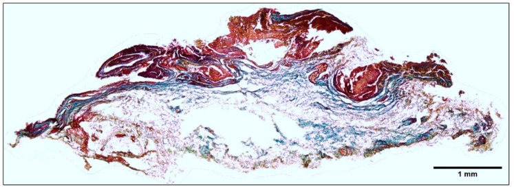
Several chronic inflammatory processes are currently being studied in relation to other systems to better understand the regulation mechanisms and identify potential therapeutic targets. A significant body of evidence supports the role of the nervous system in regulating various immunological processes. This study investigates the relationship between pterygia and the sympathetic nervous system, focusing on their interaction in the inflammatory response and fibrogenic process. Sixteen surgical specimens of primary pterygia and four conjunctival tissue samples were examined, and their morphology was analyzed using hematoxylin-eosin and Masson's trichrome stains. The gene expression of adrenergic receptors, as well as inflammatory and fibrogenic cytokines, was also assessed. Additionally, both adrenergic receptors and tyrosine hydroxylase were found to be localized within the tissues according to immunohistochemistry and immunofluorescence techniques. Increased expression of proinflammatory, fibrogenic, and adrenergic genes was observed in the pterygium compared to the healthy conjunctiva. Adrenergic receptors and tyrosine hydroxylase were localized in the basal region of the epithelium and within blood vessels, closely associated with immune cells. Neuroimmunomodulation plays a key role in the pathogenesis of pterygia by activating the sympathetic nervous system. At the intravascular level, norepinephrine promotes the migration of immune cells, thereby sustaining inflammation. Additionally, sympathetic nerve fibers located at the subepithelial level contribute to epithelial growth and the fibrosis associated with pterygia.

Keywords: fibrogenic; inflammation; neuroimmune; pterygium; response.


Conflict of interest statement

The authors declare no conflict of interest.

Figures

Figure 1
Figure 1
Panel (A): Macroscopic photograph of the pterygium. The photograph of the tissue was taken with a digital camera, using a ruler to show its length (1X). Panel (B): comparative analysis of healthy conjunctiva and pterygium. The conjunctiva (α–γ) (HEALTHY) has an epithelial layer (a), melanocytes in the basal epithelial zone (b), and a stromal layer (c) with some blood vessels (*). The pterygium (δ–ζ), with its angiomatous (γ) and fibrous (ε) histological types, shows morphological changes in its epithelial (a) and stromal (c) layers, with proliferation of blood vessels (*) and hypertrophy of goblet cells (**). Image (ζ) shows the presence of inflammatory infiltrate in the pterygium in the region near the blood vessels (*). Hematoxylin–eosin staining. Figures (α–γ) (X20)—Axiovert 40 cfl microscope (Zeiss). Figures (δ–ζ) (X10)—Axioskop 40 microscope (Zeiss). Image analysis was performed using AxioVision (Zeiss) and FIJI (Fiji Is Just Image J, GNU General Public License).
Figure 2
Figure 2
The analysis of the pterygium’s composition. The pterygium was composed of 90% collagen fibers (blue) located below the epithelial layer (red). Masson’s trichrome stain (composite image) was used field by field in the whole tissue (X10), with an Axioskop 40 microscope (Zeiss). Image analysis was performed using AxioVision (Zeiss) and FIJI (Fiji Is Just Image J, GNU General Public License).
Figure 3
Figure 3
Panel (A): Localization of tyrosine hydroxylase (TH) in pterygium tissue. Immunohistochemistry was performed using the immunoperoxidase method with diaminobenzidine (DAB) as chromogen, and images are presented as a composite from multiple fields at 10× magnification. Images (α) and (β) show higher magnifications at 40× and 100×, respectively, highlighting subepithelial localization of TH (blue arrowheads). Image (γ) shows vascular localization of TH at 40× magnification (blue arrowheads). Panel (B): Localization of the β2-adrenergic receptor (ADRB2) in pterygium. Immunohistochemistry with DAB was performed and presented as a composite image from multiple fields at 10× magnification. Image (δ) shows high magnification (100×), revealing both subepithelial (blue arrowheads) and perivascular (red arrows) localization of ADRB2. Image (ε) shows localization of the α1-adrenergic receptor (ADRA1) in perivascular smooth muscle at 100× magnification (blue arrowheads). Panel (C): Localization of TH in control conjunctival tissue. Immunohistochemistry with DAB was performed. Blue arrowheads indicate TH-positive cells in perivascular areas, suggesting the presence of sympathetic nerve fibers (40× magnification). Panel (D): Localization of ADRB2 in control conjunctival tissue. Immunohistochemistry with DAB was performed. Blue arrowheads indicate TH-positive cells in subepithelial areas, consistent with sympathetic adrenergic activity in the epithelium (40× magnification). All images were acquired using an Axioskop 40 microscope (Zeiss), and image processing and analysis were conducted using Axio-Vision (Zeiss) and FIJI (Fiji Is Just ImageJ, GNU General Public License).
Figure 4
Figure 4
The immune response in the conjunctiva and pterygium. A comparative image shows the immune response in healthy pterygium and conjunctiva. Immunofluorescence: labeling with Alexa Fluor 594® (red-orange), nuclei labeling with Hoechst 33258® (blue). Figures (A) (conjunctiva X20) and (B) (pterygium X40) show positivity for T lymphocytes (CD3), marked as *. Panel (C) (conjunctiva X20) and (D) (pterygium X40) show positivity for macrophages (CD68), marked as **. Axioskop 40 microscope (Zeiss). Image analysis was performed with AxioVision (Zeiss) and FIJI (Fiji Is Just Image J, GNU General Public License). The immune cell count in the conjunctiva and pterygium is shown. The cell count of CD3-positive T lymphocytes and CD68-positive macrophages in histological sections of healthy conjunctiva (CO) and pterygium (PT) is shown. The dots determine the cellular quantification of each patient. The mean ± standard error of the mean (SEM) is also shown; ***: p < 0.001, ****: p < 0.0001.
Figure 5
Figure 5
The neuroimmune relationship in the pterygium. The image shows the immune cell infiltrate present in the pterygium and its close relationship with TH (X20). Double immunofluorescence: labeling of macrophages and lymphocytes was performed using Alexa Fluor 594® (red-orange), labeling of TH was performed using Alexa Fluor 488® (green), and labeling of nuclei was performed using Hoechst 33258® (blue). Panel (A) shows CD3-positive T lymphocytes (*), and panel (B) shows CD68-positive macrophages (**) interacting with HT (#). Some erythrocytes were observed in the blood vessels (++). Images were taken using an Axioskop 40 microscope (Zeiss). Image analysis was performed using AxioVision (Zeiss) and FIJI (Fiji Is Just Image J, GNU General Public License).
Figure 6
Figure 6
Panel (A): The relative expression of inflammatory (IL17A and TNF-α) and fibrogenic (COL4A1 and TGFB1) genes in healthy conjunctiva (CO) and pterygium (PT). The dots indicate the gene expression of each patient. Panel (B): The relative expression of adrenergic genes (TH, ADRA1A, ADRA1B, and ADRB2) in healthy conjunctiva (CO) and pterygium (PT). The dots indicate the gene expression of each patient. Mean ± standard error of the mean (SEM) are also shown; *: p < 0.05, **: p < 0.01.

Similar articles

References

    1. Chui J., Coroneo M.T., Tat L.T., Crouch R., Wakefield D., Di Girolamo N. Ophthalmic pterygium: A stem cell disorder with premalignant features. Am. J. Pathol. 2011;178:817–827. doi: 10.1016/j.ajpath.2010.10.037. - DOI - PMC - PubMed
    1. Domdey M., Kluth M.A., Maßlo C., Ganss C., Frank M.H., Frank N.Y., Coroneo M., Cursiefen C., Notara M. Consecutive dosing of UVB irradiation induces loss of ABCB5 expression and activation of EMT and fibrosis proteins in limbal epithelial cells similar to pterygium epithelium. Stem. Cell Res. 2022;64:102936. doi: 10.1016/j.scr.2022.102936. - DOI - PMC - PubMed
    1. Malozhen S.A., Trufanov S.V., Krakhmaleva D.A. Pterygium: Etiology, pathogenesis, treatment. Vestn. Oftalmol. 2017;133:76–83. doi: 10.17116/oftalma2017133576-83. - DOI - PubMed
    1. Dushku N., John M.K., Schultz G.S., Reid T.W. Pterygia pathogenesis: Corneal invasion by matrix metalloproteinase expressing altered limbal epithelial basal cells. Arch. Ophthalmol. 2001;119:695–706. doi: 10.1001/archopht.119.5.695. - DOI - PubMed
    1. Elenkov I.J., Wilder R.L., Chrousos G.P., Vizi E.S. The Sympathetic Nerve—An Integrative Interface between Two supersystems: The brain and the Immune system. Pharmacol. Rev. 2000;52:595–638. doi: 10.1016/S0031-6997(24)01470-4. - DOI - PubMed

MeSH terms

LinkOut - more resources