. 2025 Aug 1;26(15):7423.
doi: 10.3390/ijms26157423.

Elevated IGFBP4 and Cognitive Impairment in a PTFE-Induced Mouse Model of Obstructive Sleep Apnea

Affiliations

Elevated IGFBP4 and Cognitive Impairment in a PTFE-Induced Mouse Model of Obstructive Sleep Apnea

E AlShawaf et al. Int J Mol Sci. .

Abstract

Obstructive sleep apnea (OSA) is a prevalent disorder linked to metabolic complications such as diabetes and cardiovascular disease. By fragmenting normal sleep architecture, OSA perturbs the growth hormone/insulin-like growth factor (GH/IGF) axis and alters circulating levels of IGF-binding proteins (IGFBPs). A prior clinical observation of elevated IGFBP4 in OSA patients motivated the present investigation in a controlled animal model. Building on the previously reported protocol, OSA was induced in male C57BL/6 mice (9-12 weeks old) through intralingual injection of polytetrafluoroethylene (PTFE), producing tongue hypertrophy, intermittent airway obstruction, and hypoxemia. After 8-10 weeks, the study assessed (1) hypoxia biomarkers-including HIF-1α and VEGF expression-and (2) neurobehavioral outcomes in anxiety and cognition using the open-field and novel object recognition tests. PTFE-treated mice exhibited a significant increase in circulating IGFBP4 versus both baseline and control groups. Hepatic Igfbp4 mRNA was also upregulated. Behaviorally, PTFE mice displayed heightened anxiety-like behavior and impaired novel object recognition, paralleling cognitive deficits reported in human OSA. These findings validate the PTFE-induced model as a tool for studying OSA-related hypoxia and neurocognitive dysfunction, and they underscore IGFBP4 as a promising biomarker and potential mediator of OSA's systemic effects.

Keywords: IGFBP4; OSA animal model; biomarker; hypoxia; novel object recognition; obstructive sleep apnea; open field test.


Conflict of interest statement

The authors declare no conflicts of interest.

Figures

Figure 1
Figure 1
Hypoxia induction by CoCl2, a hypoxia mimetic agent, indicated by hypoxia markers in HepG2 and the concurrent increase in IGFBP4 expression. (A) HepG2 cells showing an increase in HIF1α expression after 6 h CoCl2 treatment (mean = 1.89 ± 0.033, p = 0.015) that peaked after 48 h treatment (mean = 2.92 ± 0.19, p < 0.0001) compared with the CTRL. (B) HepG2 cells showed a significant increase in VEGF expression due to CoCl2 treatment after 24 h (mean = 3.04 ± 0.39, p = 0.007) compared with the CTRL. (C) CoCl2-induced hypoxia in HepG2 increased the expression of the IGFBP4 gene (mean = 3.67 ± 0.48, p = 0.003). A groups comparison was performed using a one-way ANOVA test, and a post hoc Dunnett test was used to correct for multiple comparisons. Statistical significance was determined with ** p-values < 0.01 and **** p-values < 0.0001.
Figure 2
Figure 2
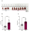
PTFE-injected tongue(s) expand significantly compared with CTRL animals. (A) Sample of extracted tongues from sacrificed animals after an 8-week follow-up period, comparing PTFE-injected mice with CTRL mice (glycerol-injected). (B) Quantification of the total tongue area shows a significant increase in the total tongue area p-value < 0.0001 of the PTFE-injected group (mean = 78.41 ± 1.6 mm2) compared with the CTRL (mean = 33.4 ± 4.7 mm2). (C) Tongue perimeter measurement shows a significant increase in the PTFE group (mean = 28.64 ± 0.81 mm) compared with the CTRL (mean = 48.34 ± 2.1 mm). Unpaired Student’s t-test was used to test statistical significance between PTFE and CTRL groups, with **** p-values < 0.0001 indicating significance.
Figure 3
Figure 3
PTFE-induced tongue expansion affects the exchange of gases. (A) A 24-h representative plot of oxygen consumption (VO2) shows a difference between the PTFE group (red) and the CTRL (black) in the amount of oxygen consumed, particularly during the dark cycle. (B) A representative plot showing a real-time recording of carbon dioxide production, with data compiled from PTFE (red) and CTRL (black). (C) Weight change and normal growth progression throughout the experiment period in all groups, with no significant difference between the groups. (D) Calculated percentage of weight gain, showing the mean percentage of weight gain throughout the study period. Weight gain percentage across groups was comparable, showing no significant difference between PTFE and CTRL.
Figure 4
Figure 4
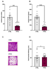
PTFE treatment induces anxiety-like behavior in animals after 8 weeks of intervention, as shown by the open-field test. (A) PTFE mice made significantly fewer entries to the central zone (18.68 ± 2.5 entry, p < 0.0001, n = 21) than CTRL mice (entry attempt = 56.7 ± 8.34, n = 10). (B) The PTFE group spent significantly less time (mean = 68.53 ± 9.9, p < 0.0001, n = 21) in the central zone than the CTRL (mean = 258.7 s ± 31.23, n = 10). (C) Representative activity trace plots of CTRL animals compared with PTFE. (D) The total distance traveled shows no difference between CTRL and PTFE (mean = 77.21 cm ± 7.5, mean = 53.41 ± 5.4, respectively). An unpaired Student’s t-test was used to test statistical significance between PTFE and CTRL groups, with *** p-values < 0.001 and **** p-values < 0.0001 indicating significance.
Figure 5
Figure 5
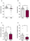
Exploring the effect of PTFE treatment on memory and learning through the novel object recognition test. (A) Discrimination index of PTFE and CTRL mice in the testing phase, showing a significantly reduced cognitive ability in the PTFE group (mean = −0.043 ± 0.083, p = 0.016, n = 21) compared with the CTRL (mean = 0.24 ± 0.046, n = 13). (B) Recognition index in the testing phase, showing a significantly lower value in the PTFE group (mean = 0.478 ± 0.041, p = 0.0161, n = 21) compared with the CTRL (mean = 0.62 ± 0.023, n = 13). (C) Total distance traveled by animals from both groups is comparable with no significant difference (CTRL = 11.6 ± 1.4, PTFE = 9.15 ± 0.98, p = 0.15). (D) Novel object investigation attempts by the PTFE group (mean = 8.09 ± 1.5 times) were significantly less than attempts by the CTRL (mean = 15.69 ± 3.7, p = 0.037). An unpaired Student’s t-test was used to test statistical significance between PTFE and CTRL groups, with * p-values < 0.05 indicating significance.
Figure 6
Figure 6
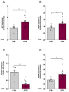
Intermittent hypoxia induces the expression of IGFBP4 at the gene level in the liver. Tongue-induced expansion increased the gene expression of hypoxia markers; (A) HIF1α in PTFE = 1.72 ± 0.28, p = 0.044 compared with CTRL = 1.08 ± 0.08. (B) VEGF expression level in PTFE = 1.24 ± 0.09, p = 0.042 compared with CTRL = 0.903 ± 0.08. (C) IGFBP3 gene expression significantly declined in PTFE = 0.29 ± 0.1, p = 0.025 compared with the CTRL = 0.98 ± 0.29. (D) IGFBP4 gene expression increased significantly in PTFE = 1.51 ± 0.15, p = 0.048 compared with CTRL = 0.93 ± 0.09. An unpaired Student’s t-test was used to test statistical significance between PTFE and CTRL groups, with * p-values < 0.05 indicating significance.
Figure 7
Figure 7
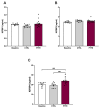
Significant increase in circulating IGFBP4 with intermittent hypoxia. To evaluate the effect of hypoxia on IGFBPs, we quantified circulation. (A) IGFPB1 showing no difference in plasma IGFBP1 levels between baseline (mean = 1.39 ± 0.02 ng/mL) and post-intervention (PTFE = 1.4 ± 0.07 ng/mL, CTRL = 1.34 ± 0.04 ng/mL). (B) No significant change in IGFBP3 levels before (mean = 2.4 ± 0.09 ng/mL) and after intervention (PTFE = 2.54 ± 0.03 ng/mL, CTRL = 2.58 ± 0.02 ng/mL). (C) Circulating IGFBP4 levels showed a significant increase in PTFE group (mean = 23.65 ± 0.67 ng/mL) compared with CTRL (mean = 19.55 ± 1.3 ng/mL) and baseline (mean = 20.2 ± 0.98 ng/mL). Groups comparison was performed with a one-way ANOVA test, and a post hoc Dunnett test was used to correct for multiple comparisons. Statistical significance was determined with ** p-values < 0.01.

Similar articles

References

    1. Lévy P., Kohler M., McNicholas W.T., Barbé F., McEvoy R.D., Somers V.K., Lavie L., Pépin J.L. Obstructive sleep apnoea syndrome. Nat. Rev. Dis. Primers. 2015;1:15015. doi: 10.1038/nrdp.2015.15. - DOI - PubMed
    1. Senaratna C.V., Perret J.L., Lodge C.J., Lowe A.J., Campbell B.E., Matheson M.C., Hamilton G.S., Dharmage S.C. Prevalence of obstructive sleep apnea in the general population: A systematic review. Sleep Med. Rev. 2017;34:70–81. doi: 10.1016/j.smrv.2016.07.002. - DOI - PubMed
    1. Chen X., Wang R., Zee P., Lutsey P.L., Javaheri S., Alcántara C., Jackson C.L., Williams M.A., Redline S. Racial/Ethnic Differences in Sleep Disturbances: The Multi-Ethnic Study of Atherosclerosis (MESA) Sleep. 2015;38:877–888. doi: 10.5665/sleep.4732. - DOI - PMC - PubMed
    1. Jehan S., Zizi F., Pandi-Perumal S.R., Wall S., Auguste E., Myers A.K., Jean-Louis G., McFarlane S.I. Obstructive Sleep Apnea and Obesity: Implications for Public Health. Sleep Med. Disord. Int. J. 2017;1:00019. doi: 10.15406/smdij.2017.01.00019. - DOI - PMC - PubMed
    1. Theorell-Haglöw J., Miller C.B., Bartlett D.J., Yee B.J., Openshaw H.D., Grunstein R.R. Gender differences in obstructive sleep apnoea, insomnia and restless legs syndrome in adults—What do we know? A clinical update. Sleep Med. Rev. 2018;38:28–38. doi: 10.1016/j.smrv.2017.03.003. - DOI - PubMed

MeSH terms

Substances

LinkOut - more resources