. 2025 Aug 1;26(15):7434.
doi: 10.3390/ijms26157434.

Resveratrol Impairs Insulin Signaling in Hepatic Cells via Activation of PKC and PTP1B Pathways

Affiliations

Resveratrol Impairs Insulin Signaling in Hepatic Cells via Activation of PKC and PTP1B Pathways

Karla D Hernández-González et al. Int J Mol Sci. .

Abstract

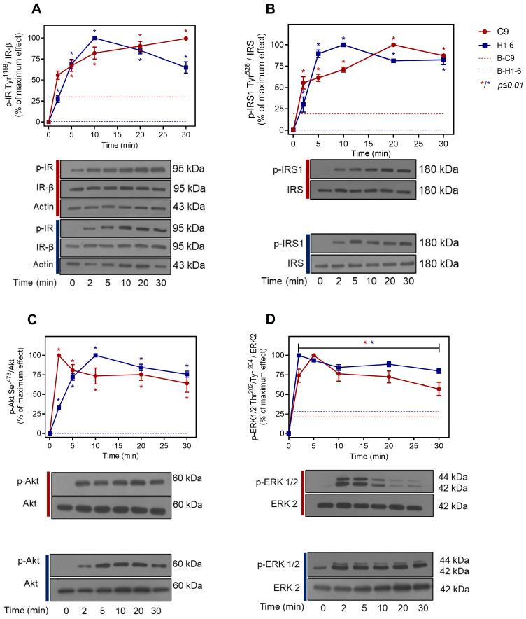
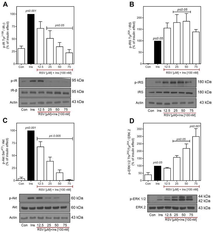
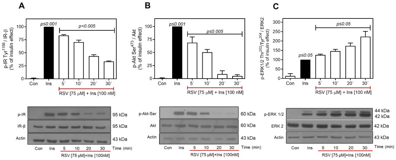
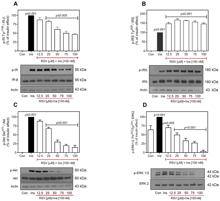
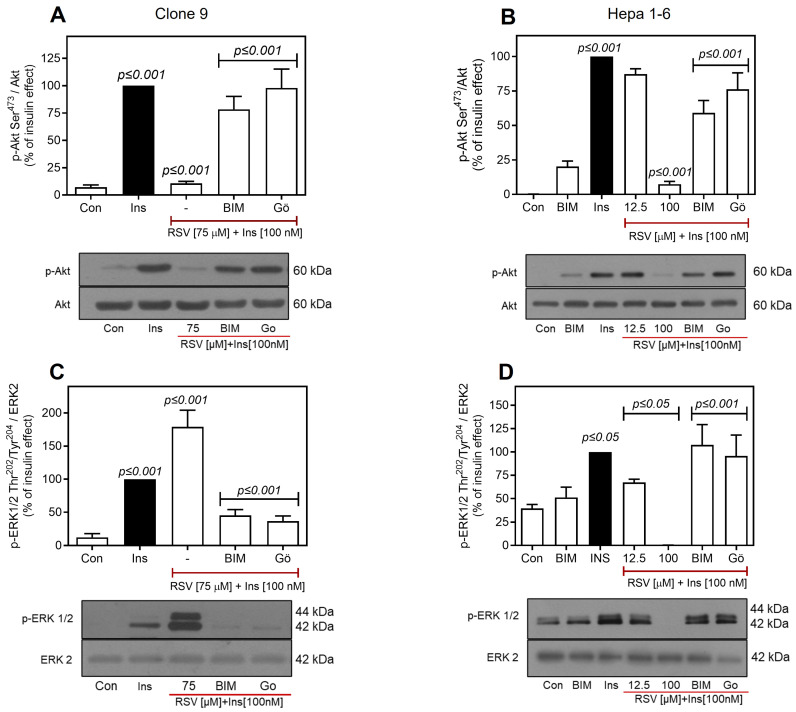
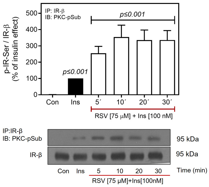
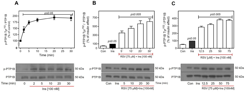
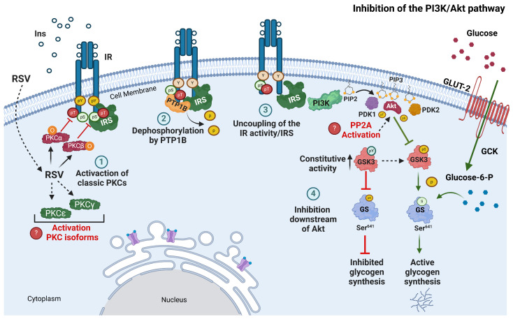
Resveratrol (RSV), a polyphenol found in a variety of berries and wines, is known for its anti-inflammatory, anticancer, and antioxidant properties. It has been suggested that RSV may play a role in the regulation of metabolic disorders, including diabetes and insulin resistance. However, in recent years, it has been reported to completely inhibit Akt kinase function in liver cells. Akt is a central protein involved in the metabolic function of insulin and is regulated by the phosphatidylinositol-3-kinase (PI3K) pathway. In this study, we examined the effect of RSV on insulin-induced insulin receptor (IR) phosphorylation and proteins involved in the PI3K/Akt pathway in a hepatic cell model, clone 9 (C9), and in hepatoma cells, Hepa 1-6 (H1-6). In both cell lines, RSV inhibited tyrosine phosphorylation of IR and insulin-induced activation of Akt. We also evaluated the effect of RSV on the activation of protein tyrosine phosphatase 1B (PTP1B), which is associated with IR dephosphorylation, and found that RSV increased PTP1B-Tyr152 phosphorylation in a time- and concentration-dependent manner. Furthermore, we found that the protein kinase C (PKC) inhibitors BIM and Gö6976 prevented the inhibition of Akt phosphorylation by RSV and increased the phosphorylation of Ser/Thr residues in IR, suggesting that PKC is involved in the inhibition of the insulin pathway by RSV. Thus, classical PKC isoforms impair the PI3K/Akt pathway at the IR and GSK3 and GS downstream levels; however, IRS-Tyr632 phosphorylation remains unaffected. These results suggest that RSV can lead to insulin resistance by activating PTP1B and PKC, consequently affecting glucose homeostasis in hepatic cells.

Keywords: hepatic cells; insulin receptor; insulin resistance; protein kinase C; protein tyrosine phosphatase; resveratrol.


Conflict of interest statement

The authors declare that the research was conducted without any commercial or financial relationships that could be construed as a potential conflict of interest.

Figures

Figure 1
Figure 1
Activation of the insulin pathway in hepatic cells. C9 and Hepa 1-6 cells were stimulated with 100 nM insulin for the indicated times. Total cell lysates were separated by SDS-PAGE and analyzed by immunoblotting using anti-p-IR-Tyr1158 (A), anti-p-IRS-1-Tyr628 (B), anti-p-Akt-Ser473 (C), and anti-p-ERK1/2-Thr202/Tyr204 (D), as described in Materials and Methods. Data represent the mean ± S.E.M. of five independent experiments, and the panels below show representative blots. Western blots were also probed for total IR, IRS, Akt, ERK, and actin, all showing equal loading. Vertical lines represent the S.E.M. values. * p-value ≤ 0.01 vs. basal phosphorylation (AD). C9, clone 9 cells (red circles); H1-6, Hepa 1-6 cells (blue squares); B-C9, basal phosphorylation of C9 (red dotted line); B-H1-6, basal phosphorylation of H1-6 (blue dotted line).
Figure 2
Figure 2
Effect of RSV on insulin-induced phosphorylation of IR/IRS/Akt and ERK in C9 cells. C9 cells were pretreated with different concentrations of RSV (12.5–75 µM as indicated) and stimulated with 100 nM insulin for an additional 10 min. Under all experimental conditions, untreated cells were used as controls. Total cell lysates were separated by SD-PAGE and analyzed by immunoblotting with anti-p-IR-Tyr1158 (A), anti-p-IRS-1-Tyr628 (B), anti-p-Akt-Ser473 (C), or anti-p-ERK1/2-Thr202/Tyr204 (D), as described in Materials and Methods. Data represent the mean ± S.E.M. of five independent experiments, and the panels below show representative immunoblots. Western blots were also probed for total IR, IRS, Akt, and ERK1/2, which showed equal loading. Vertical lines represent the S.E.M. The p-value indicates: Ins vs. control (AD); RSV [12.5–75 µM] + Ins [100 nM] vs. Ins (AD).
Figure 3
Figure 3
RSV affects time-dependent insulin-induced phosphorylation of IR/Akt. C9 cells were pretreated with RSV (75 µM) for the indicated times and then stimulated with 100 nM insulin for an additional 10 min. Under all experimental conditions, untreated cells were used as controls. Total cell lysates were separated by SD-PAGE and analyzed by immunoblotting with anti-p-IR-Tyr1158 (A), anti-p-Akt-Ser473 (B), or anti-p-ERK1/2-Thr202/Tyr204 (C), as described in Materials and Methods. Western blots were also probed for total IR, Akt, and ERK1/2 showing equal loading. Data represent the mean ± S.E.M. of five to six individual experiments, and the panels below show representative immunoblots. Vertical lines represent the S.E.M. The p-value indicates Ins vs. control (AC); RSV (5–30 min) + Ins [100 nM] vs. Ins (AC).
Figure 4
Figure 4
Effect of RSV on insulin-induced phosphorylation of IR/IRS/Akt and ERK in Hepa 1-6 cells. Hepa 1-6 cells were pretreated with different concentrations of RSV (12.5–100 µM as indicated) and then stimulated with 100 nM insulin for an additional 10 min. Under all experimental conditions, untreated cells were used as controls. Total cell lysates were separated by SD-PAGE and analyzed by immunoblotting with anti-p-IR-Tyr1158 (A), anti-p-IRS-1-Tyr628 (B), anti-p-Akt-Ser473 (C), or anti-p-ERK1/2-Thr202/Tyr204 (D), as described in Materials and Methods. Western blots were also probed for total IR, IRS, Akt, and ERK1/2 showing equal loading. Data represent the mean ± S.E.M. of five to six individual experiments, and the panels below show representative immunoblots. Vertical lines represent the S.E.M. The p-value indicates Ins vs. control (AD); RSV [12.5–100 µM] + Ins [100 nM] vs. Ins (AD).
Figure 5
Figure 5
RSV negatively regulates the insulin pathway downstream of Akt. C9 cells were pretreated with different concentrations of RSV (12.5–75 µM as indicated) and then stimulated with 100 nM insulin for an additional 10 min. Under all experimental conditions, untreated cells were used as controls. Total cell lysates were separated by SD-PAGE and analyzed by immunoblotting with anti-p-Akt-Thr308 (A) or anti-p-GSK3α/β-Ser21/9 (B) and anti-p-GS-Ser641 (C), as described in Materials and Methods. Representative blots of Akt, GSK3, and GS phosphorylation are shown (D). * The same blot was used in Figure 2C, as it corresponds to the same experiment. Western blots were also probed for total Akt showing equal loading. Data represent the mean ± S.E.M. of five to six individual experiments, and the panels below show representative immunoblots. Vertical lines represent the S.E.M. The p-value indicates Ins vs. control (AD); RSV [12.5–75 µM] + Ins [100 nM] vs. Ins (AD).
Figure 6
Figure 6
Role of PKC in RSV-mediated regulation of insulin signaling. C9 and Hepa 1-6 cells were pretreated with or without 1 µM BIM or 100 nM Gö6976 for 30 min, and then treated with different concentrations of RSV (as indicated) for 30 min. Finally, the cells were stimulated with 100 nM insulin for an additional 10 min. Under all experimental conditions, untreated cells were used as controls. Total cell lysates were separated by SD-PAGE and analyzed by immunoblotting with anti-p-Akt-Ser473 (A,B) or anti-p-ERK1/2-Thr202/Tyr204 (C,D), as described in Materials and Methods. Western blots were also probed for total Akt and ERK2, which showed equal loading. Data represent the mean ± S.E.M. of five to six individual experiments, and the panels below show representative immunoblots. Vertical lines represent the S.E.M. The p-value indicates Ins vs. control (AD); RSV [75 µM] + Ins [100 nM] (A,C) or RSV [12.5 and 100 µM] + Ins [100 nM] vs. Ins (B,D); BIM or Gö vs. RSV [75 µM] + Ins [100 nM] (A,C) or RSV [100 µM] + Ins [100 nM] (B,D).
Figure 7
Figure 7
RSV promotes IR-Ser-phosphorylation through PKC activation. C9 cells were treated with or without RSV (75 µM) for 5–30 min and then stimulated with 100 nM insulin for an additional 10 min. Under all experimental conditions, untreated cells were used as controls. Total cell lysates were immunoprecipitated with an IR antibody before SDS-PAGE analysis and immunoblotted with anti-phospho-Ser-PKC substrate antibody, as described in Materials and Methods. Western blots were also probed for total IR-β showing equal loading. Data represent the mean ± S.E.M. of five to six individual experiments, and the panels below show representative immunoblots. Vertical lines represent the S.E.M. The p-value indicates Ins vs. control; RSV (5–30 min) + Ins [100 nM] vs. Ins.
Figure 8
Figure 8
Activation of PTP1B by RSV. C9 cells were treated with 100 nM insulin for 2–30 min (A), pretreated with RSV (75 µM) for 5–30 min, followed by stimulation with 100 nM insulin for an additional 10 min (B), or pretreated with different concentrations of RSV (12.5–75 µM as indicated) for 30 min, and then stimulated with 100 nM insulin for an additional 10 min (C). Under all experimental conditions, untreated cells were used as controls. Total cell lysates were separated by SD-PAGE and analyzed by immunoblotting with anti-p-PTP1B-Tyr152 (AC), as described in Materials and Methods. Western blots were also probed for total PTP1B showing equal loading. Data represent the mean ± S.E.M. of five to six individual experiments, and the panels below show representative immunoblots. Vertical lines represent the S.E.M. The p-value indicates Ins vs. control (baseline in 0 min) (A); Ins vs. Con (B,C); RSV (5–30 min) + Ins [100 nM] vs. Ins (B,C).
Figure 9
Figure 9
Model of desensitization of insulin signaling pathway by RSV in C9 cells. When hepatic cells are treated with RSV, classical PKC isoforms are activated, which in turn interact with the insulin receptor (IR) and phosphorylate it on serine residues (Ser). Subsequently, PTP1B dephosphorylates the receptor and causes uncoupling with IRS, impairing insulin actions, such as hepatic glycogen synthesis. The numbered circles indicate the process of inhibition of the PI3K/Akt pathway by RSV: (1) RSV activates classic isoforms of PKC that phosphorylate Ser/Thr residues in IR; (2) previous phosphorylation of Ser/Thr in IR facilitates the interaction of PTP1B with IR and dephosphorylates it; (3) inhibition of IR causes uncoupling of IRS; and (4) downstream of the PI3K/Akt pathway is inhibited. Green arrows indicate normal insulin regulation, and red arrows indicate the inhibition of RSV-regulated processes. The diagram indicates hypothetical processes in red letters that could contribute to the negative regulation of the PI3K/Akt pathway, such as the activation of other PKC isoforms and the PP2A phosphatase. Ins, insulin; GCK, glucokinase; pSer, serine phosphorylation; pThr, threonine phosphorylation; pTyr, tyrosine phosphorylation; RSV, resveratrol. Figure created using BioRender.com (2024).

Similar articles

References

    1. Li Z., Chen X., Liu G., Li J., Zhang J., Cao Y., Miao J. Antioxidant Activity and Mechanism of Resveratrol and Polydatin Isolated from Mulberry (Morus Alba L.) Molecules. 2021;26:7574. doi: 10.3390/molecules26247574. - DOI - PMC - PubMed
    1. Akinwumi B., Bordun K.-A., Anderson H. Biological Activities of Stilbenoids. Int. J. Mol. Sci. 2018;19:792. doi: 10.3390/ijms19030792. - DOI - PMC - PubMed
    1. Salehi B., Mishra A.P., Nigam M., Sener B., Kilic M., Sharifi-Rad M., Fokou P.V.T., Martins N., Sharifi-Rad J. Resveratrol: A Double-Edged Sword in Health Benefits. Biomedicines. 2018;6:91. doi: 10.3390/biomedicines6030091. - DOI - PMC - PubMed
    1. Tong W., Chen X., Song X., Chen Y., Jia R., Zou Y., Li L., Yin L., He C., Liang X., et al. Resveratrol Inhibits Lps-Induced Inflammation through Suppressing the Signaling Cascades of Tlr4-Nf-κb/Mapks/Irf3. Exp. Ther. Med. 2020;19:1824–1834. doi: 10.3892/etm.2019.8396. - DOI - PMC - PubMed
    1. Kursvietiene L., Kopustinskiene D.M., Staneviciene I., Mongirdiene A., Kubová K., Masteikova R., Bernatoniene J. Anti-Cancer Properties of Resveratrol: A Focus on Its Impact on Mitochondrial Functions. Antioxidants. 2023;12:2056. doi: 10.3390/antiox12122056. - DOI - PMC - PubMed

LinkOut - more resources