Osteogenic Differentiation of Mesenchymal Stem Cells Induced by Geometric Mechanotransductive 3D-Printed Poly-(L)-Lactic Acid Matrices
- PMID: 40806620
- PMCID: PMC12347987
- DOI: 10.3390/ijms26157494
Osteogenic Differentiation of Mesenchymal Stem Cells Induced by Geometric Mechanotransductive 3D-Printed Poly-(L)-Lactic Acid Matrices
Abstract
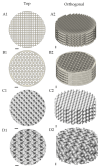
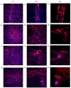
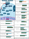
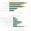
Bone-related defects present a key challenge in orthopaedics. The current gold standard, autografts, poses significant limitations, such as donor site morbidity, limited supply, and poor morphological adaptability. This study investigates the potential of scaffold geometry to induce osteogenic differentiation of human adipose-derived stem cells (hADSCs) through mechanotransduction, without the use of chemical inducers. Four distinct poly-(L)-lactic acid (PLA) scaffold architectures-Traditional Cross (Tc), Triangle (T), Diamond (D), and Gyroid (G)-were fabricated using fused filament fabrication (FFF) 3D printing. hADSCs were cultured on these scaffolds, and their response was evaluated utilising an alkaline phosphatase (ALP) assay, immunofluorescence, and extensive proteomic analyses. The results showed the D scaffold to have the highest ALP activity, followed by Tc. Proteomics results showed that more than 1200 proteins were identified in each scaffold with unique proteins expressed in each scaffold, respectively Tc-204, T-194, D-244, and G-216. Bioinformatics analysis revealed structures with complex curvature to have an increased expression of proteins involved in mid- to late-stage osteogenesis signalling and differentiation pathways, while the Tc scaffold induced an increased expression of signalling and differentiation pathways pertaining to angiogenesis and early osteogenesis.
Keywords: adipose-derived stem cells (ADSCs); bone tissue engineering (BTE); mechanotransduction.
Conflict of interest statement
The authors declare no conflicts of interest.
Figures


Similar articles
-
Mineralized osteoblast-derived exosomes and 3D-printed ceramic-based scaffolds for enhanced bone healing: A preclinical exploration.Acta Biomater. 2025 Jun 15;200:686-702. doi: 10.1016/j.actbio.2025.05.051. Epub 2025 May 21. Acta Biomater. 2025. PMID: 40409510
-
3D-printed nano-hydroxyapatite/poly(lactic-co-glycolic acid) scaffolds with adipose-derived mesenchymal stem cells enhance bone regeneration in rat model of bone defects.J Biomater Appl. 2025 Aug;40(2):284-296. doi: 10.1177/08853282251332050. Epub 2025 Apr 3. J Biomater Appl. 2025. PMID: 40179420
-
Odontogenic/osteogenic differentiation of dental pulp stem cells on a Biodentine-coated polymer nanofibers.Biomed Eng Online. 2025 Jul 17;24(1):91. doi: 10.1186/s12938-025-01421-5. Biomed Eng Online. 2025. PMID: 40676689 Free PMC article.
-
4D printing of smart scaffolds for bone regeneration: a systematic review.Biomed Mater. 2024 Dec 25;20(1). doi: 10.1088/1748-605X/ad8f80. Biomed Mater. 2024. PMID: 39504649
-
Use of 3D-printed polylactic acid/bioceramic composite scaffolds for bone tissue engineering in preclinical in vivo studies: A systematic review.Acta Biomater. 2023 Sep 15;168:1-21. doi: 10.1016/j.actbio.2023.07.013. Epub 2023 Jul 15. Acta Biomater. 2023. PMID: 37454707
References
-
- Abdelaziz A.G., Nageh H., Abdo S.M., Abdalla M.S., Amer A.A., Abdal-Hay A., Barhoum A. A Review of 3D Polymeric Scaffolds for Bone Tissue Engineering: Principles, Fabrication Techniques, Immunomodulatory Roles, and Challenges. Bioengineering. 2023;10:204. doi: 10.3390/bioengineering10020204. - DOI - PMC - PubMed
MeSH terms
Substances
LinkOut - more resources
Full Text Sources

