. 2025 Aug 5;26(15):7558.
doi: 10.3390/ijms26157558.

An Italian Single-Center Genomic Surveillance Study: Two-Year Analysis of SARS-CoV-2 Spike Protein Mutations

Affiliations

An Italian Single-Center Genomic Surveillance Study: Two-Year Analysis of SARS-CoV-2 Spike Protein Mutations

Riccardo Cecchetto et al. Int J Mol Sci. .

Abstract

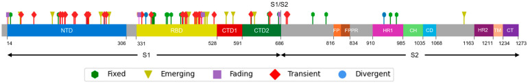
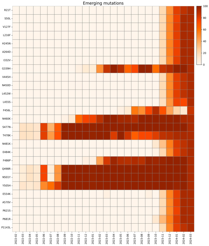
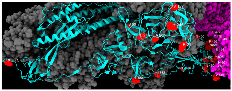
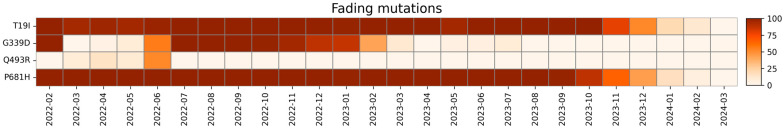
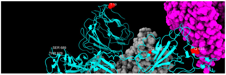
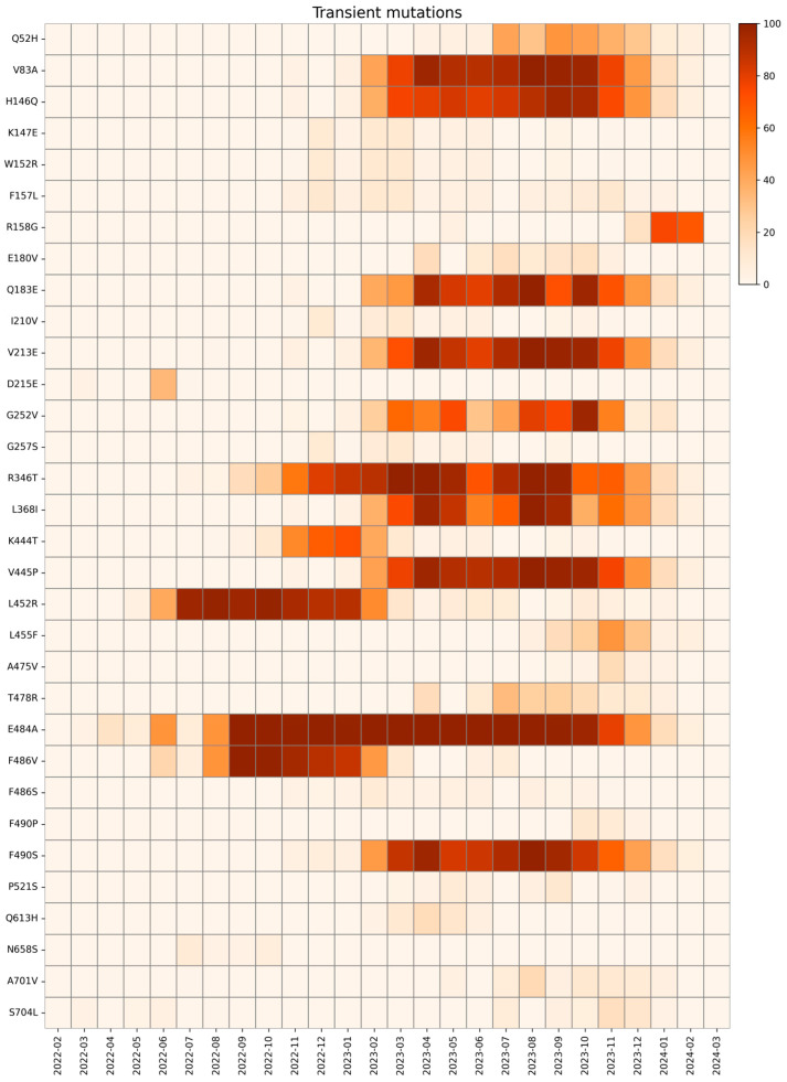
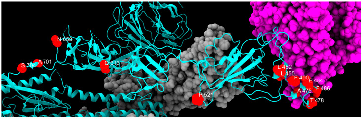
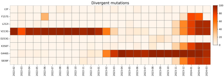
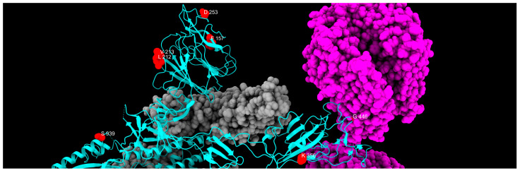
The repeated occurrence of SARS-CoV-2 variants, largely driven by virus-host interactions, was and will remain a public health concern. Spike protein mutations shaped viral infectivity, transmissibility, and immune escape. From February 2022 to April 2024, a local genomic surveillance program in Verona, Italy, was conducted on 1333 SARS-CoV-2-positive nasopharyngeal swabs via next generation full-length genome sequencing. Spike protein mutations were classified based on their prevalence over time. Mutations were grouped into five categories: fixed, emerging, fading, transient, and divergent. Notably, some divergent mutations displayed a "Lazarus effect," disappearing and later reappearing in new lineages, indicating potential adaptive advantages in specific genomic contexts. This two-year surveillance study highlights the dynamic nature of spike protein mutations and their role in SARS-CoV-2 evolution. The findings underscore the need for ongoing mutation-focused genomic monitoring to detect early signals of variant emergence, especially among mutations previously considered disadvantageous. Such efforts are critical for driving public health responses and guiding future vaccine and therapeutic strategies.

Keywords: SARS-CoV-2; genomic surveillance; mutational analysis; spike protein; whole genome sequencing.


Conflict of interest statement

The authors declare no conflicts of interest.

Figures

Figure 1
Figure 1
Mutation distribution along the S protein. Each mutation is colored differently according to the following classification: green, fixed; yellow, emerging; violet, fading; red, transient; and blue, divergent mutations. S1: Subunit 1; S2: Subunit 2; NTD: N-Terminal Domain; RBD: Receptor Binding Domain; CTD1: C-terminal domain 1; and CTD2: C-terminal domain 2; FP: fusion peptide; FPPR: fusion-peptide proximal region; HR1: heptad repeat 1; CH: central helix; CD: connector domain; HR2: heptad repeat 2; TM: transmembrane segment; CT: cytoplasmic tail.
Figure 2
Figure 2
Three-dimensional structure of spike protein, with all mutations described in this study highlighted in different colors: green, fixed; yellow, emerging; violet, fading; red, transient; and blue, divergent mutations. Cyan, gray, and magenta are used to represent one chain of the protein, the other spike chains of the trimer, and the ACE2 receptor, respectively.
Figure 3
Figure 3
Spearman’s correlation heatmap matrix for all substitutions, grouped by the classification given in this study. The colored boxes highlight the specific correlations between substitutions belonging to the same group: green, fixed; yellow, emerging; violet, fading; red, transient; and blue, divergent mutations.
Figure 4
Figure 4
Plot of Spearman’s correlation index of the global trend compared to that observed in this study for each mutation. Almost all mutations display a positive correlation index, indicating that the trend of substitution considered in this study mirrors the global one. Panels (AD) each show 22 substitutions, ordered according to their amino acid position along the S protein.
Figure 5
Figure 5
Distribution of fixed mutation sites, colored in red, along one chain of S protein, colored in cyan. Gray and magenta represent the other spike chains of the trimer and the ACE2 receptor, respectively.
Figure 6
Figure 6
Heatmap with the relative monthly prevalence of the emerging mutations in the period from February 2022 to March 2024.
Figure 7
Figure 7
Distribution of emerging mutation sites, colored in red, along one chain of S protein, colored in cyan. Gray and magenta represent the other spike chains of the trimer and the ACE2 receptor, respectively.
Figure 8
Figure 8
Heatmap with the relative monthly prevalence of the fading mutations in the period from February 2022 to March 2024.
Figure 9
Figure 9
Distribution of fading mutation sites, colored in red, along one chain of S protein, colored in cyan, with the exception of P681H, which is absent from the PDB model and is limited to a side loop. Gray and magenta represent the other spike chains of the trimer and the ACE2 receptor, respectively.
Figure 10
Figure 10
Heatmap with the relative monthly prevalence of transient mutations in the period from February 2022 to March 2024.
Figure 11
Figure 11
Distribution of transient mutation sites, colored in red, along one chain of S protein, colored in cyan. Gray and magenta represent the other spike chains of the trimer and the ACE2 receptor, respectively.
Figure 12
Figure 12
Heatmap with relative monthly prevalence of divergent mutations in the period from February 2022 to March 2024.
Figure 13
Figure 13
Distribution of divergent mutation sites, colored in red, along one chain of the S protein, colored in cyan. Gray and magenta represent the other spike chains of the trimer and the ACE2 receptor, respectively.
Figure 14
Figure 14
Venn diagram showing the co-occurrence of mutation involved, either positively or negatively, in immune evasion and ACE2 binding.

Similar articles

References

    1. Lazarevic I., Pravica V., Miljanovic D., Cupic M. Immune Evasion of SARS-CoV-2 Emerging Variants: What Have We Learnt So Far? Viruses. 2021;13:1192. doi: 10.3390/v13071192. - DOI - PMC - PubMed
    1. Flores-Vega V.R., Monroy-Molina J.V., Jiménez-Hernández L.E., Torres A.G., Santos-Preciado J.I., Rosales-Reyes R. SARS-CoV-2: Evolution and Emergence of New Viral Variants. Viruses. 2022;14:653. doi: 10.3390/v14040653. - DOI - PMC - PubMed
    1. De Castro E., Hulo C., Masson P., Auchincloss A., Bridge A., Le Mercier P. ViralZone 2024 Provides Higher-Resolution Images and Advanced Virus-Specific Resources. Nucleic Acids Res. 2024;52:D817–D821. doi: 10.1093/nar/gkad946. - DOI - PMC - PubMed
    1. Naqvi A.A.T., Fatima K., Mohammad T., Fatima U., Singh I.K., Singh A., Atif S.M., Hariprasad G., Hasan G.M., Hassan M.d.I. Insights into SARS-CoV-2 Genome, Structure, Evolution, Pathogenesis and Therapies: Structural Genomics Approach. Biochim. Et Biophys. Acta (BBA) Mol. Basis Dis. 2020;1866:165878. doi: 10.1016/j.bbadis.2020.165878. - DOI - PMC - PubMed
    1. Lu R., Zhao X., Li J., Niu P., Yang B., Wu H., Wang W., Song H., Huang B., Zhu N., et al. Genomic Characterisation and Epidemiology of 2019 Novel Coronavirus: Implications for Virus Origins and Receptor Binding. Lancet. 2020;395:565–574. doi: 10.1016/S0140-6736(20)30251-8. - DOI - PMC - PubMed

LinkOut - more resources