. 2025 Aug 5;26(15):7560.
doi: 10.3390/ijms26157560.

Lycopene Inhibits PRRSV Replication by Suppressing ROS Production

Affiliations

Lycopene Inhibits PRRSV Replication by Suppressing ROS Production

Ying-Xian Ma et al. Int J Mol Sci. .

Abstract

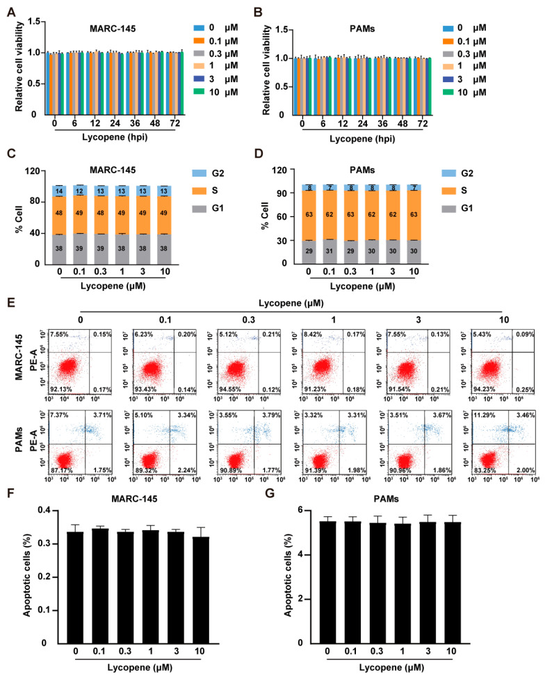
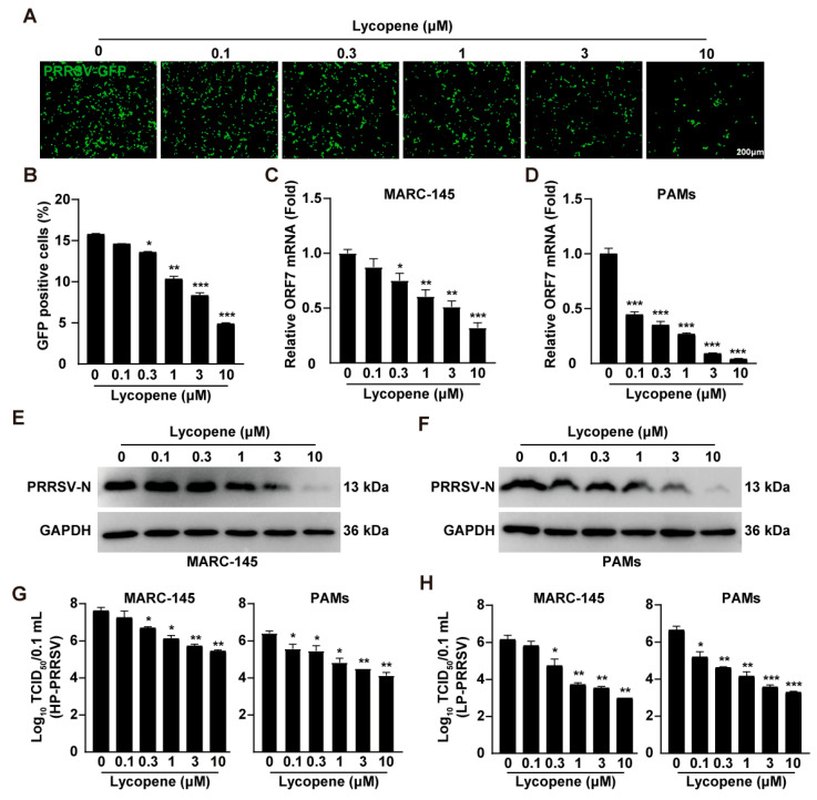
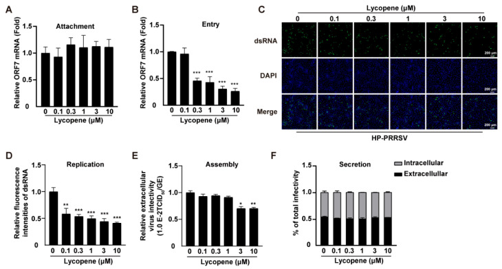
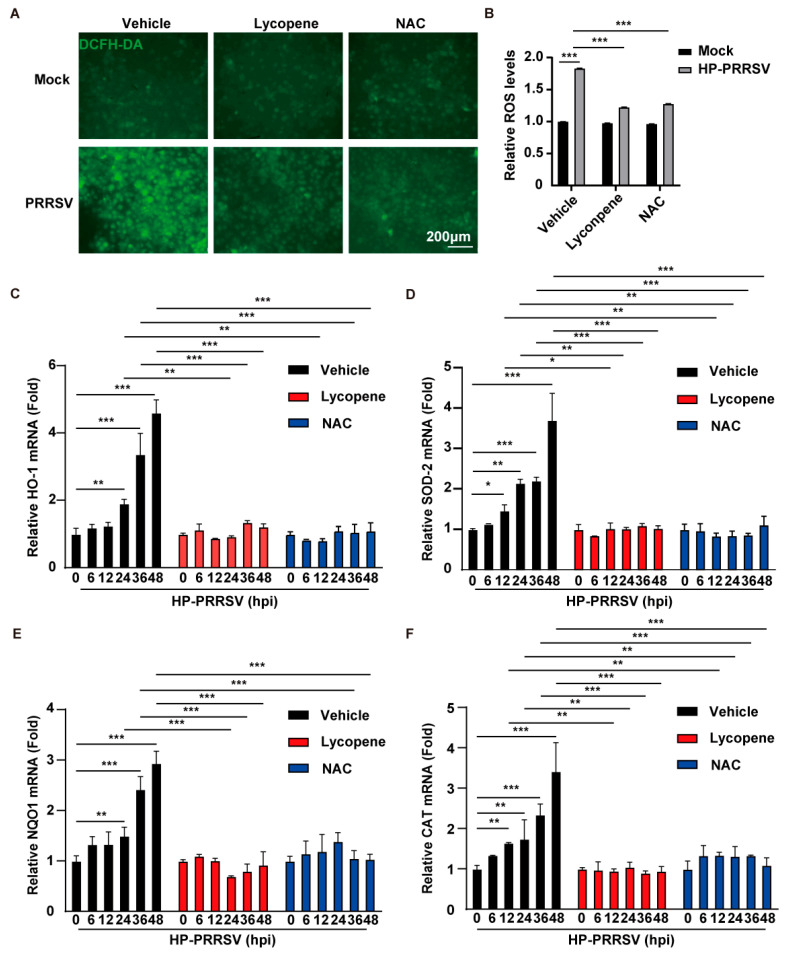
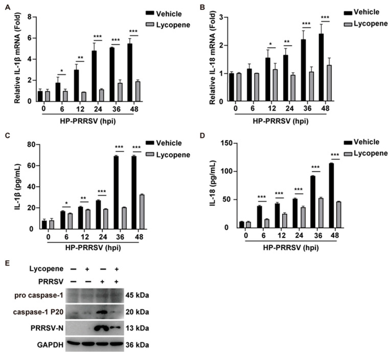
Porcine reproductive and respiratory syndrome virus (PRRSV), an enveloped single-stranded positive-sense RNA virus, poses a significant threat to global swine production. Despite the availability of modified live virus and inactivated vaccines, their limited efficacy and safety concerns highlight the urgent need for novel antiviral therapeutics. This study aimed to investigate the molecular mechanisms by which lycopene inhibits PRRSV replication. Initial assessments confirmed that lycopene did not adversely affect cellular viability, cell cycle progression, or apoptosis. Using fluorescence microscopy, flow cytometry, immunoblotting, quantitative real-time PCR (qRT-PCR), and viral titration assays, lycopene was shown to exhibit potent antiviral activity against PRRSV. Mechanistic studies revealed that lycopene suppresses reactive oxygen species (ROS) production, which is critical for PRRSV proliferation. Additionally, lycopene attenuated PRRSV-induced inflammatory responses, as demonstrated by immunoblotting, ELISA, and qRT-PCR assays. These findings suggest that lycopene inhibits PRRSV replication by modulating ROS levels and mitigating inflammation, offering a promising avenue for the development of antiviral therapeutics. This study provides new insights and strategies for combating PRRSV infections, emphasizing the potential of lycopene as a safe and effective antiviral agent.

Keywords: PRRSV; ROS; inflammation; lycopene.


Conflict of interest statement

The authors declare that they have no competing interests.

Figures

Figure 1
Figure 1
Effect of lycopene on cell viability, cell cycle, and apoptosis. (A,B) MARC-145 (A) and PAM (B) cells were treated with lycopene (0–10 μM) for 0–72 h. Cell viability was assessed with CCK-8 assay. (C,D) MARC-145 (C) and PAM (D) cells were treated with lycopene (0–10 μM) for 36 h, and the cell cycle was assessed by flow cytometry following Hoechst 33, 342 staining. (EG) MARC-145 and PAMs were treated with lycopene (0–10 μM) for 36 h. Cell apoptosis was evaluated by flow cytometry using Annexin V-FITC and PI staining (E). Panels (F,G) show the percentage of cell death.
Figure 2
Figure 2
Lycopene inhibits PRRSV replication in vitro. (A) MARC-145 cells were infected with PRRSV-GFP (MOI = 10) and treated with lycopene (0–10 μM) for 48 h. Virus replication was observed using fluorescence microscopy. (B) Flow cytometry analysis of GFP-positive cells from panel (A). *** p < 0.001; ** p < 0.01; * p < 0.05 (the p value is calculated vs. control). (C,D) MARC-145 and PAMs were infected with HP-PRRSV (MOI = 1) and treated with lycopene (0–10 μM) for 48 h. The mRNA level of PRRSV ORF7 was analyzed by qRT-PCR. *** p < 0.001; ** p < 0.01; * p < 0.05 (the p value is calculated vs. control). (E,F) MARC-145 (E) and PAMs (F) were treated as in C, and PRRSV-N and GAPDH were detected by immunoblotting. (G,H) MARC-145 and PAMs were infected with HP-PRRSV (MOI = 10) and LP-PRRSV (MOI = 10) and treated with lycopene (0–10 μM) for 48 h. Virus titers were determined by TCID50 assay *** p < 0.001; ** p < 0.01; * p < 0.05 (the p value is calculated vs. control).
Figure 3
Figure 3
Lycopene disrupts PRRSV entry, replication, and assembly. (A) HP-PRRSV (MOI = 10) was incubated with MARC-145 cells at 4 °C for 1 h in the presence of lycopene (0–10 μM). After washing the cells three times with cold PBS, total RNA was extracted and reverse-transcribed into cDNA. Virus attachment was evaluated by the qRT-PCR analysis of PRRSV ORF7 mRNA. (B) HP-PRRSV (MOI = 10) was incubated with MARC-145 cells at 4 °C for 1 h, followed by incubation at 37 °C for 2 h in a medium containing lycopene (0–10 μM). After washing the cells three times with PBS, total RNA was extracted and reverse-transcribed into cDNA. Virus entry was evaluated by qRT-PCR analysis of PRRSV ORF7 mRNA. *** p < 0.001 (the p value is calculated vs. control). (C) MARC-145 cells were infected with HP-PRRSV (MOI = 10) and treated with lycopene (0–10 μM) for 36 h. Virus replication was evaluated by dsRNA staining. (D) Quantification of relative fluorescence intensity of dsRNA from panel (C). *** p < 0.001; ** p < 0.01 (the p value is calculated vs. control). (E) MARC-145 cells were infected with HP-PRRSV (MOI = 10) and treated with lycopene (0–10 μM) for 36 h. Virus assembly efficiency in the supernatant was determined by comparing the infection titer (TCID50/mL) with the total PRRSV genome equivalent (ORF7). ** p < 0.01; * p < 0.05 (the p value is calculated vs. control). (F) MARC-145 cells were infected with HP-PRRSV (MOI = 10) and treated with lycopene (0–10 μM) for 36 h. Virus secretion efficiency was measured by the ratio of intracellular and extracellular infectivity to total infectivity.
Figure 4
Figure 4
Lycopene inhibits PRRSV-induced ROS. (A) MARC-145 cells were infected with HP-PRRSV (MOI = 1) and treated with 10 μM of lycopene or 10 mM of NAC for 24 h. The ROS levels in the cells were determined by DCFH-DA staining. Scale bar: 200 μm. (B) Quantification of relative ROS levels from panel (A) using ImageJ (v1.8.0.345). *** p < 0.001 (the p value is calculated vs. control). (CF) MARC-145 cells were infected with HP-PRRSV (MOI = 1) and treated with 10 μM of lycopene or 10 mM of NAC for 24 h. qRT-PCR was used to analyze the mRNA expression levels of the HO-1, SOD-2, NQO1, and CAT genes in the cells. *** p < 0.001; ** p < 0.01; * p < 0.05 (the p value is calculated vs. control).
Figure 5
Figure 5
Lycopene inhibits PRRSV-induced inflammatory response. (A,B) PAM cells were infected with HP-PRRSV (MOI = 1) and treated with 10 μM of lycopene for 0–48 h. qRT-PCR analysis was conducted to evaluate the mRNA expression levels of IL-1β and IL-18 in the cells. *** p < 0.001; ** p < 0.01; * p < 0.05 (the p value is calculated vs. control). (C,D) PAM cells were infected with HP-PRRSV (MOI = 1) and treated with 10 μM of lycopene for 0–48 h. ELISA was used to measure the secretion levels of IL-1β and IL-18 in the cell supernatant. *** p < 0.001; ** p < 0.01; * p < 0.05 (the p value is calculated vs. control). (E) PAM cells were infected with PRRSV (MOI = 1) and treated with 10 μM of lycopene for 48 h. Immunoblotting was performed to analyze the levels of pro-caspase-1, caspase-1 P20, and GAPDH.

Similar articles

References

    1. Lunney J.K., Fang Y., Ladinig A., Chen N., Li Y., Rowland B., Renukaradhya G.J. Porcine Reproductive and Respiratory Syndrome Virus (PRRSV): Pathogenesis and Interaction with the Immune System. Annu. Rev. Anim. Biosci. 2016;4:129–154. doi: 10.1146/annurev-animal-022114-111025. - DOI - PubMed
    1. Collins J.E., Benfield D.A., Christianson W.T., Harris L., Hennings J.C., Shaw D.P., Goyal S.M., McCullough S., Morrison R.B., Joo H.S., et al. Isolation of swine infertility and respiratory syndrome virus (isolate ATCC VR-2332) in North America and experimental reproduction of the disease in gnotobiotic pigs. J. Vet. Diagn. Invest. 1992;4:117–126. doi: 10.1177/104063879200400201. - DOI - PubMed
    1. Baron T., Albina E., Leforban Y., Madec F., Guilmoto H., Plana Duran J., Vannier P. Report on the first outbreaks of the porcine reproductive and respiratory syndrome (PRRS) in France. Diagnosis and viral isolation. Ann. Rech. Vet. 1992;23:161–166. - PubMed
    1. Conzelmann K.K., Visser N., Van Woensel P., Thiel H.J. Molecular characterization of porcine reproductive and respiratory syndrome virus, a member of the arterivirus group. Virology. 1993;193:329–339. doi: 10.1006/viro.1993.1129. - DOI - PMC - PubMed
    1. Snijder E.J., Meulenberg J.J. The molecular biology of arteriviruses. Pt 5J. Gen. Virol. 1998;79:961–979. doi: 10.1099/0022-1317-79-5-961. - DOI - PubMed

LinkOut - more resources