. 2025 Aug 5;26(15):7584.
doi: 10.3390/ijms26157584.

Structural Insights and Calcium-Switching Mechanism of Fasciola hepatica Calcium-Binding Protein FhCaBP4

Affiliations

Structural Insights and Calcium-Switching Mechanism of Fasciola hepatica Calcium-Binding Protein FhCaBP4

Byeongmin Shin et al. Int J Mol Sci. .

Abstract

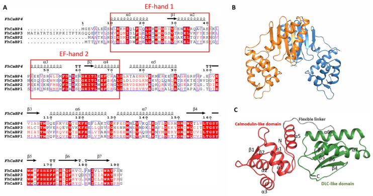
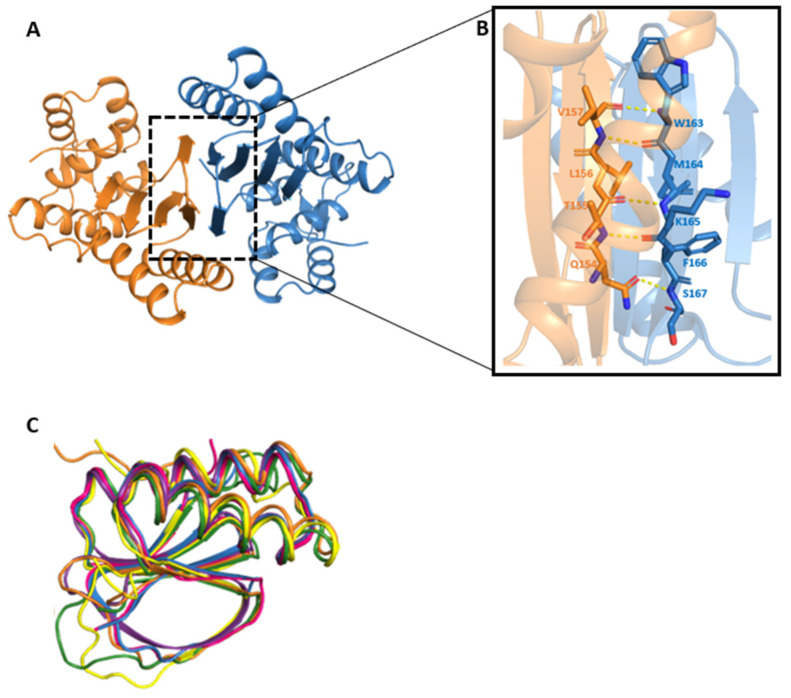
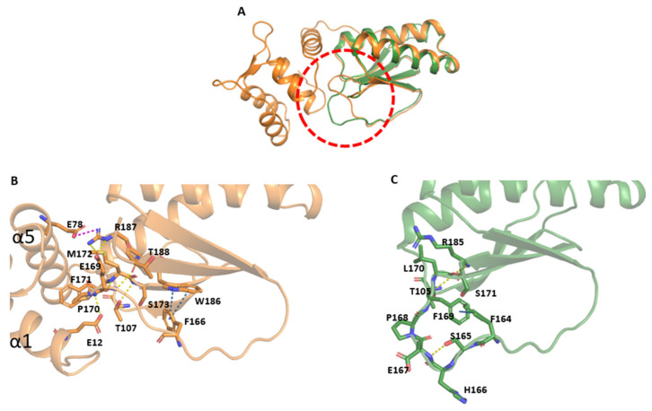
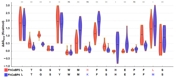
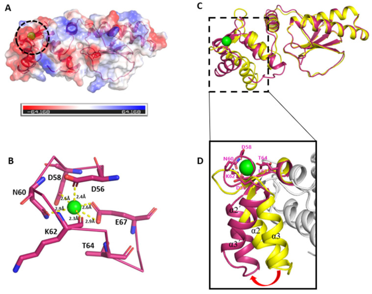
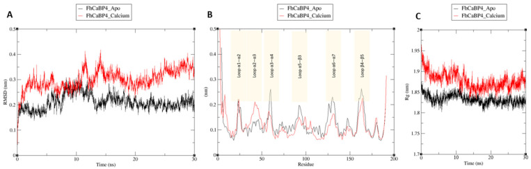
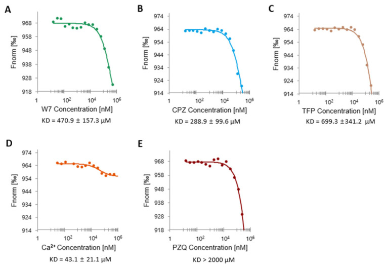
Fasciola hepatica remains a global health and economic concern, and treatment still relies heavily on triclabendazole. At the parasite-host interface, F. hepatica calcium-binding proteins (FhCaBPs) have a unique EF-hand/DLC-like domain fusion found only in trematodes. This makes it a parasite-specific target for small compounds and vaccinations. To enable novel therapeutic strategies, we report the first elevated-resolution structure of a full-length FhCaBP4. The apo structure was determined at 1.93 Å resolution, revealing a homodimer architecture that integrates an N-terminal, calmodulin-like, EF-hand pair with a C-terminal dynein light chain (DLC)-like domain. Structure-guided in silico mutagenesis identified a flexible, 16-residue β4-β5 loop (LTGSYWMKFSHEPFMS) with an FSHEPF core that demonstrates greater energetic variability than its FhCaBP2 counterpart, likely explaining the distinct ligand-binding profiles of these paralogs. Molecular dynamics simulations and AlphaFold3 modeling suggest that EF-hand 2 acts as the primary calcium-binding site, with calcium coordination inducing partial rigidification and modest expansion of the protein structure. Microscale thermophoresis confirmed calcium as the major ligand, while calmodulin antagonists bound with lower affinity and praziquantel demonstrated no interaction. Thermal shift assays revealed calcium-dependent stabilization and a merger of biphasic unfolding transitions. These results suggest that FhCaBP4 functions as a calcium-responsive signaling hub, with an allosterically coupled EF-hand-DLC interface that could serve as a structurally tractable platform for drug targeting in trematodes.

Keywords: DLC-like domain; EF-hand; Fasciola hepatica; FhCaBP4; calcium; homodimer.


Conflict of interest statement

The authors declare no conflicts of interest.

Figures

Figure 1
Figure 1
Overall structure of FhCaBP4. (A) Multiple-sequence alignment of the four CaBP paralogs (FhCaBP1–4). Positions identical in ≥70% of the sequences are shaded red, while conservative substitutions appear in pink. Secondary-structure elements assigned from the present FhCaBP4 structure are displayed above the alignment (black cylinders, α-helices α1–α6; black arrows, β-strands β1–β7). The two EF-hand calcium-binding loops are boxed in red. (B) Dimeric structure of FhCaBP4. The dimeric structure is shown as a cartoon diagram showing with orange and blue, respectively. (C) Monomeric structure of FhCaBP4. The calmodulin-like domain is shown in red, the dynein light chain (DLC)-like domain in green, and the intervening Gly-rich linker in gray. Structural elements are labeled sequentially (α1–α7, β1–β6). N- and C-termini are labeled.
Figure 2
Figure 2
Dimeric structure of the FhCaBP4 and its DLC-like domain (A) Dimeric structure of the FhCaBP4. One monomer is colored in orange and the other in sky-blue, represented as a cartoon model. The dimeric interface area is represented by a black dashed rectangle. (B) Close-up view of the dimeric interface. Yellow dashed lines represent hydrogen bonds of the primary chains, and the interacting residues are represented as a stick model. (C) Superimposition of the DLC-like domains. The DLC-like domain backbone models of Drosophila melanogaster (PDB3BRI), Toxoplasma gondii (PDB3RJS), Saccharomyces cerevisiae (PDB4DS1), Clonorchis sinensis (PDB5X2D), Fasciola hepatica (PDB5FX0), and FhCaBP4 are shown in blue, pink, purple, yellow, green, and orange, respectively.
Figure 3
Figure 3
Structural comparison of FhCaBP4 and the DLC-like domain of FhCaBP2. (A) Superposition of FhCaBP4 (orange) and FhCaBP2 DLC-like domain (PDB ID: 5FXO; green) displayed in cartoon representation. The flexible loop region is highlighted by a red dashed circle. (B) Zoomed-in view of the flexible loop region in the FhCaBP4 structure. Residues involved in hydrogen bonding are connected by yellow dashed lines, salt–bridge interactions by pink dashed lines, and π–π interactions by blue dashed lines. All interacting side chains are shown in stick representation. (C) Zoomed-in view of the corresponding loop region in the DLC-like domain of FhCaBP2. Hydrogen bonds are indicated by yellow dashed lines and π–π interactions by blue dashed lines. All interacting side chains are rendered as sticks.
Figure 4
Figure 4
Mutational scanning of the loop in FhCaBP2 (red) and FhCaBP4 (blue). Violin plots show ΔΔGbind distributions when each position in the loop sequence was substituted with the 19 other amino acids in FhCaBP2 and FhCaBP4. Horizontal lines indicate medians. Asterisks indicate significance by two-sided Wilcoxon rank-sum test (*** p < 0.001; ** p < 0.01; * p < 0.05; ns, not significant).
Figure 5
Figure 5
The predicted calcium-bound structure of FhCaBP4 and its conformational changes. (A) Electrostatic surfaces of the predicted calcium-bound FhCaBP4. The calcium-binding site is represented by a black dashed circle. (B) Clos-up view of the predicted calcium-binding site. The green sphere represents calcium ion. The yellow dashed lines represent the interaction with calcium ions, and residues that interact with calcium are represented as a stick model. (C) Superstition of apo-FhCaBP4 (yellow) with an AlphaFold3-predicted calcium-bound model of its calmodulin-like domain (warm pink). The calcium ion at the EF-hand 2 is depicted as a green sphere and the deep view is represented as a black dashed rectangle. (D) Close-up view of the moving region. The red arrow highlights an outward rotation of helices α2 and α3 upon calcium binding. The residues that interact with calcium ions are represented as a stick model.
Figure 6
Figure 6
Molecular dynamics analysis of FhCaBP4 apo and calcium-bound form. (A) Time-dependent root mean square deviation (RMSD) trajectories for FhCaBP4 apo (black line) and calcium-bound FhCaBP4 (red line) showing backbone stability over 30 ns. (B) Residue-wise root mean square fluctuation (RMSF) profiles for the same trajectories. Shaded boxes highlight six loop regions (Loop α1–α2, Loop α2–α3, Loop α3–α4, Loop α5–β3, Loop α6–α7, and Loop β4–β5) where differences in flexibility are most pronounced between apo (black) and calcium-bound (red) forms. (C) Radius of gyration (Rg) as a function of simulation time, illustrating that the calcium-bound form (red) maintains a higher average Rg than the apo form (black).
Figure 7
Figure 7
Microscale thermophoresis (MST) analysis of FhCaBP4 binding to chemical ligands. Binding isotherms were generated by plotting normalized fluorescence (Fnorm, %) as a function of increasing ligand concentrations. Each assay was conducted with 100 nM labeled FhCaBP4 and a serial dilution of the corresponding ligand (0.015–500 μM): (A) W7, Kd = 470.9 ± 157.3 μM; (B) CPZ, Kd = 288.9 ± 99.6 μM; (C) TFP, Kd = 699.3 ± 341.2 μM; (D) Calcium, Kd = 43.1 ± 21.1 μM; (E) PZQ, Kd > 2000 μM. All data represent mean values from at least three independent experiments, each using sixteen capillaries per condition. Dissociation constants (Kd) were calculated by using MO Affinity Analysis software v2.1.3. with a nonlinear quadratic binding model. Values are reported as mean ± SD. μM, micromolar; nM, nanomolar.

Similar articles

References

    1. Caravedo M.A., Cabada M.M. Human fascioliasis: Current epidemiological status and strategies for diagnosis, treatment, and control. Res. Rep. Trop. Med. 2020;11:149–158. doi: 10.2147/RRTM.S237461. - DOI - PMC - PubMed
    1. Mehmood K., Zhang H., Sabir A.J., Abbas R.Z., Ijaz M., Durrani A.Z., Saleem M.H., Rehman M.U., Iqbal M.K., Wang Y. A review on epidemiology, global prevalence and economical losses of fasciolosis in ruminants. Microb. Pathog. 2017;109:253–262. doi: 10.1016/j.micpath.2017.06.006. - DOI - PubMed
    1. Kelley J.M., Elliott T.P., Beddoe T., Anderson G., Skuce P., Spithill T.W. Current threat of triclabendazole resistance in Fasciola hepatica. Trends Parasitol. 2016;32:458–469. doi: 10.1016/j.pt.2016.03.002. - DOI - PubMed
    1. Choi Y.-J., Rosa B.A., Fernandez-Baca M.V., Ore R.A., Martin J., Ortiz P., Hoban C., Cabada M.M., Mitreva M. Independent origins and non-parallel selection signatures of triclabendazole resistance in Fasciola hepatica. Nat. Commun. 2025;16:2996. doi: 10.1038/s41467-025-57796-5. - DOI - PMC - PubMed
    1. Beesley N.J., Cwiklinski K., Allen K., Hoyle R.C., Spithill T.W., La Course E.J., Williams D.J., Paterson S., Hodgkinson J.E. A major locus confers triclabendazole resistance in Fasciola hepatica and shows dominant inheritance. PLoS Pathog. 2023;19:e1011081. doi: 10.1371/journal.ppat.1011081. - DOI - PMC - PubMed

LinkOut - more resources