

Transcriptomic Profiling of Mouse Mesenchymal Stem Cells Exposed to Metal-Based Nanoparticles
- PMID: 40806712
- PMCID: PMC12347195
- DOI: 10.3390/ijms26157583
Transcriptomic Profiling of Mouse Mesenchymal Stem Cells Exposed to Metal-Based Nanoparticles
Abstract
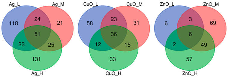
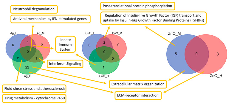
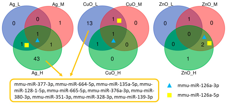
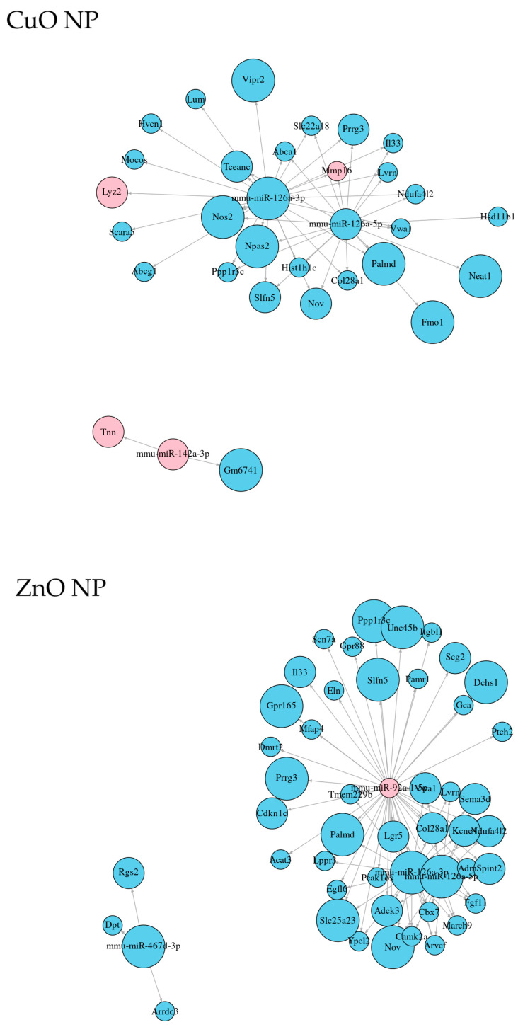
Mesenchymal stem cells (MSCs), i.e., adult stem cells with immunomodulatory and secretory properties, contribute to tissue growth and regeneration, including healing processes. Some metal nanoparticles (NPs) are known to exhibit antimicrobial activity and may further potentiate tissue healing. We studied the effect of Ag, CuO, and ZnO NPs after in vitro exposure of mouse MSCs at the transcriptional level in order to reveal the potential toxicity as well as modulation of other processes that may modify the activity of MSCs. mRNA-miRNA interactions were further investigated to explore the epigenetic regulation of gene expression. All the tested NPs mediated immunomodulatory effects on MSCs, generation of extracellular vesicles, inhibition of osteogenesis, and enhancement of adipogenesis. Ag NPs exhibited the most pronounced response; they impacted the expression of the highest number of mRNAs, including those encoding interferon-γ-stimulated genes and genes involved in drug metabolism/cytochrome P450 activity, suggesting a response to the potential toxicity of Ag NPs (oxidative stress). Highly interacting MiR-126 was upregulated by all NPs, while downregulation of MiR-92a was observed after the ZnO NP treatment only, and both effects might be associated with the improvement of MSCs' healing potency. Overall, our results demonstrate positive effects of NPs on MSCs, although increased oxidative stress caused by Ag NPs may limit the therapeutical potential of the combined MSC+NP treatment.
Keywords: in vitro exposure; mouse mesenchymal stem cells; nanoparticles; whole-genome expression analysis of mRNA and miRNA.
Conflict of interest statement
The authors declare no conflicts of interest.
Figures

Similar articles
-
Prescription of Controlled Substances: Benefits and Risks.2025 Jul 6. In: StatPearls [Internet]. Treasure Island (FL): StatPearls Publishing; 2025 Jan–. 2025 Jul 6. In: StatPearls [Internet]. Treasure Island (FL): StatPearls Publishing; 2025 Jan–. PMID: 30726003 Free Books & Documents.
-
Systemic treatments for metastatic cutaneous melanoma.Cochrane Database Syst Rev. 2018 Feb 6;2(2):CD011123. doi: 10.1002/14651858.CD011123.pub2. Cochrane Database Syst Rev. 2018. PMID: 29405038 Free PMC article.
-
The Black Book of Psychotropic Dosing and Monitoring.Psychopharmacol Bull. 2024 Jul 8;54(3):8-59. Psychopharmacol Bull. 2024. PMID: 38993656 Free PMC article. Review.
-
Skin Wound Healing: The Impact of Treatment with Antimicrobial Nanoparticles and Mesenchymal Stem Cells.J Xenobiot. 2025 Jul 18;15(4):119. doi: 10.3390/jox15040119. J Xenobiot. 2025. PMID: 40700166 Free PMC article.
-
Measures implemented in the school setting to contain the COVID-19 pandemic.Cochrane Database Syst Rev. 2022 Jan 17;1(1):CD015029. doi: 10.1002/14651858.CD015029. Cochrane Database Syst Rev. 2022. Update in: Cochrane Database Syst Rev. 2024 May 2;5:CD015029. doi: 10.1002/14651858.CD015029.pub2. PMID: 35037252 Free PMC article. Updated.
References
LinkOut - more resources
Full Text Sources
Miscellaneous
