

ArfGAP with Dual Pleckstrin Homology Domains 2 Promotes Hypertrophy of Cultured Neonatal Cardiomyocytes
- PMID: 40806715
- PMCID: PMC12347555
- DOI: 10.3390/ijms26157588
ArfGAP with Dual Pleckstrin Homology Domains 2 Promotes Hypertrophy of Cultured Neonatal Cardiomyocytes
Abstract
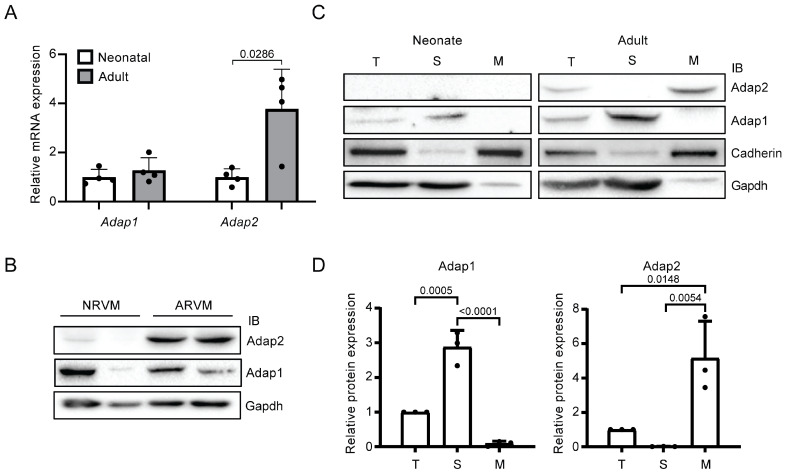
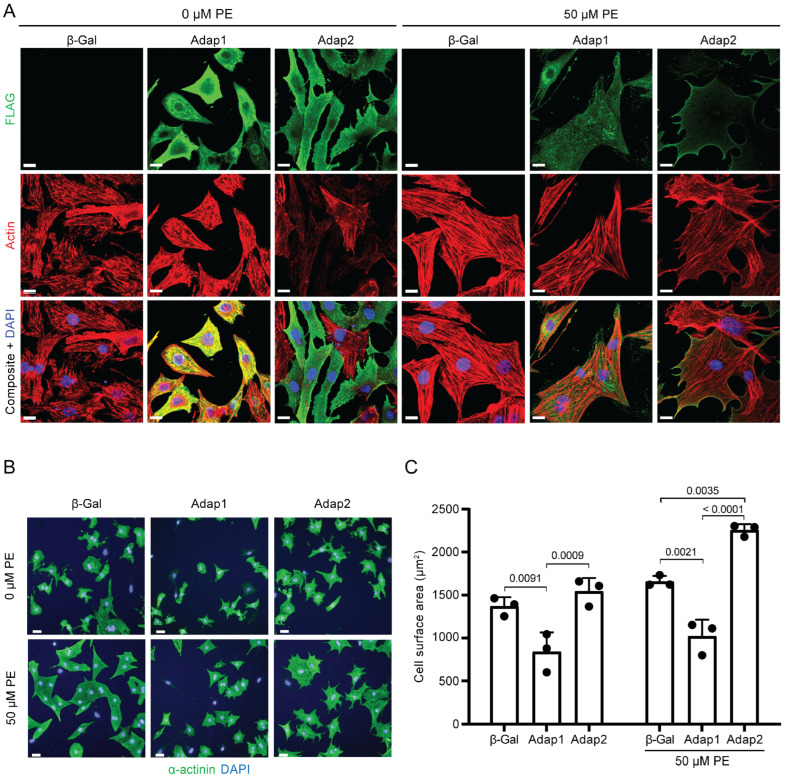
Cardiomyocyte hypertrophy is regulated by several factors, including the ADP-ribosylation factor (Arf) family of small G proteins, among others. For instance, ArfGAP with dual pleckstrin homology domains 1 (Adap1) exerts an anti-hypertrophic effect in cultured cardiomyocytes. Its homologous protein, Adap2, is also expressed in the heart but its role remains elusive. To elucidate its function, we investigated the effects of adenoviral-mediated overexpression of Adap2 in cultured neonatal rat ventricular myocytes under both basal and pro-hypertrophic conditions, employing a range of microscopy and biochemical techniques. Despite minimal detection in neonatal rat hearts, Adap2 was found to be well expressed in adult rat hearts, being predominantly localized at the membrane fraction. In contrast to Adap1, overexpression of Adap2 provokes the robust accumulation of β1-integrin at the cellular surface of cultured cardiomyocytes. Interestingly, overexpressed Adap2 relocalizes at the sarcolemma and increases the size of cardiomyocytes upon phenylephrine stimulation, despite attenuating Erk1/2 phosphorylation and Nppa gene expression. Under these same conditions, cardiomyocytes overexpressing Adap2 also express higher level of detyrosinated tubulin, a marker of hypertrophic response. These findings provide new insights into the pro-hypertrophic function of Adap2 in cardiomyocytes.
Keywords: Adap1; Adap2; Centaurin-α; cardiomyocyte; hypertrophy; tubulin detyrosination.
Conflict of interest statement
The authors declare that the research was conducted in the absence of any commercial or financial relationships that could be construed as a potential conflict of interest.
Figures


Similar articles
-
ADAP1 limits neonatal cardiomyocyte hypertrophy by reducing integrin cell surface expression.Sci Rep. 2018 Sep 11;8(1):13605. doi: 10.1038/s41598-018-31784-w. Sci Rep. 2018. PMID: 30206251 Free PMC article.
-
Cardiac-specific overexpression of PRMT5 exacerbates pressure overload-induced hypertrophy and heart failure.J Biomed Sci. 2025 Jul 6;32(1):61. doi: 10.1186/s12929-025-01162-6. J Biomed Sci. 2025. PMID: 40619438 Free PMC article.
-
Artemisinin Attenuates Isoproterenol-induced Cardiac Hypertrophy via the ERK1/2 and p38 MAPK Signaling Pathways.Curr Mol Pharmacol. 2024;17:e18761429244886. doi: 10.2174/0118761429244886230927070818. Curr Mol Pharmacol. 2024. PMID: 38258596
-
Systemic pharmacological treatments for chronic plaque psoriasis: a network meta-analysis.Cochrane Database Syst Rev. 2017 Dec 22;12(12):CD011535. doi: 10.1002/14651858.CD011535.pub2. Cochrane Database Syst Rev. 2017. Update in: Cochrane Database Syst Rev. 2020 Jan 9;1:CD011535. doi: 10.1002/14651858.CD011535.pub3. PMID: 29271481 Free PMC article. Updated.
-
Maternal and neonatal outcomes of elective induction of labor.Evid Rep Technol Assess (Full Rep). 2009 Mar;(176):1-257. Evid Rep Technol Assess (Full Rep). 2009. PMID: 19408970 Free PMC article.
References
-
- Venkateswarlu K., Oatey P.B., Tavare J.M., Jackson T.R., Cullen P.J. Identification of centaurin-alpha1 as a potential in vivo phosphatidylinositol 3,4,5-trisphosphate-binding protein that is functionally homologous to the yeast ADP-ribosylation factor (ARF) GTPase-activating protein, Gcs1. Biochem. J. 1999;340:359–363. doi: 10.1042/bj3400359. - DOI - PMC - PubMed
-
- Hammonds-Odie L.P., Jackson T.R., Profit A.A., Blader I.J., Turck C.W., Prestwich G.D., Theibert A.B. Identification and cloning of centaurin-alpha. A novel phosphatidylinositol 3,4,5-trisphosphate-binding protein from rat brain. J. Biol. Chem. 1996;271:18859–18868. doi: 10.1074/jbc.271.31.18859. - DOI - PMC - PubMed
MeSH terms
Substances
Grants and funding
LinkOut - more resources
Full Text Sources
Miscellaneous
