

Epigenetic Alterations in Age-Related Macular Degeneration: Mechanisms and Implications
- PMID: 40806731
- PMCID: PMC12347735
- DOI: 10.3390/ijms26157601
Epigenetic Alterations in Age-Related Macular Degeneration: Mechanisms and Implications
Abstract
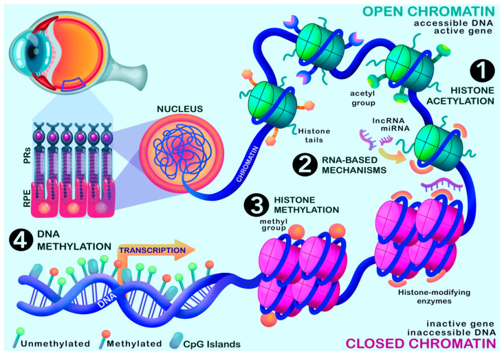
Age-related macular degeneration (AMD) is one of the leading causes of irreversible vision loss among the elderly, and is influenced by a combination of genetic and environmental risk factors. While genetic associations in AMD are well-established, the molecular mechanisms underlying disease onset and progression remain poorly understood. A growing body of evidence suggests that epigenetic modifications may serve as a potential missing link regulating gene-environment interactions. This review incorporates recent findings on DNA methylation, including both hypermethylation and hypomethylation patterns affecting genes such as silent mating type information regulation 2 homolog 1 (SIRT1), glutathione S-transferase isoform (GSTM), and SKI proto-oncogene (SKI), which may influence key pathophysiological drivers of AMD. We also examine histone modification patterns, chromatin accessibility, the status of long non-coding RNAs (lncRNAs) in AMD pathogenesis and in regulating pathways pertinent to the pathophysiology of the disease. While the field of ocular epigenetics remains in its infancy, accumulating evidence to date points to a burgeoning role for epigenetic regulation in AMD, pre-clinical studies have yielded promising findings for the prospect of epigenetics as a future therapeutic avenue.
Keywords: DNA methylation; age-related macular degeneration; chromatin accessibility; epigenetics; histone acetylation; histone methylation; histone modifications; histone variants; long noncoding RNA.
Conflict of interest statement
The authors declare no conflicts of interest.
Figures


Similar articles
-
A systematic review of epigenetic interplay in kidney diseases: Crosstalk between long noncoding RNAs and methylation, acetylation of chromatin and histone.Biomed Pharmacother. 2024 Jul;176:116922. doi: 10.1016/j.biopha.2024.116922. Epub 2024 Jun 12. Biomed Pharmacother. 2024. PMID: 38870627
-
[Epigenetics' implication in autism spectrum disorders: A review].Encephale. 2017 Aug;43(4):374-381. doi: 10.1016/j.encep.2016.07.007. Epub 2016 Sep 28. Encephale. 2017. PMID: 27692350 French.
-
Surgery for cataracts in people with age-related macular degeneration.Cochrane Database Syst Rev. 2017 Feb 16;2(2):CD006757. doi: 10.1002/14651858.CD006757.pub4. Cochrane Database Syst Rev. 2017. PMID: 28206671 Free PMC article.
-
Prescription of Controlled Substances: Benefits and Risks.2025 Jul 6. In: StatPearls [Internet]. Treasure Island (FL): StatPearls Publishing; 2025 Jan–. 2025 Jul 6. In: StatPearls [Internet]. Treasure Island (FL): StatPearls Publishing; 2025 Jan–. PMID: 30726003 Free Books & Documents.
-
Anti-vascular endothelial growth factor biosimilars for neovascular age-related macular degeneration.Cochrane Database Syst Rev. 2024 Jun 3;6(6):CD015804. doi: 10.1002/14651858.CD015804.pub2. Cochrane Database Syst Rev. 2024. PMID: 38829176 Free PMC article.
References
-
- Smith W., Assink J., Klein R., Mitchell P., Klaver C.C., Klein B.E., Hofman A., Jensen S., Wang J.J., de Jong P.T.V.M., et al. Risk factors for age-related macular degeneration: Pooled findings from three continents. Ophthalmology. 2001;108:697–704. doi: 10.1016/S0161-6420(00)00580-7. - DOI - PubMed
-
- Wong W.L., Su X., Li X., Cheung C.M.G., Klein R., Cheng C.-Y., Wong T.Y. Global prevalence of age-related macular degeneration and disease burden projection for 2020 and 2040: A systematic review and meta-analysis. Lancet Glob. Health. 2014;2:e106–e116. doi: 10.1016/S2214-109X(13)70145-1. - DOI - PubMed
Publication types
MeSH terms
Substances
Grants and funding
LinkOut - more resources
Full Text Sources
Medical
Research Materials
