. 2025 Aug 6;26(15):7612.
doi: 10.3390/ijms26157612.

AMPK-Targeting Effects of (-)-Epicatechin Gallate from Hibiscus sabdariffa Linne Leaves on Dual Modulation of Hepatic Lipid Accumulation and Glycogen Synthesis in an In Vitro Oleic Acid Model

Affiliations

AMPK-Targeting Effects of (-)-Epicatechin Gallate from Hibiscus sabdariffa Linne Leaves on Dual Modulation of Hepatic Lipid Accumulation and Glycogen Synthesis in an In Vitro Oleic Acid Model

Hui-Hsuan Lin et al. Int J Mol Sci. .

Abstract

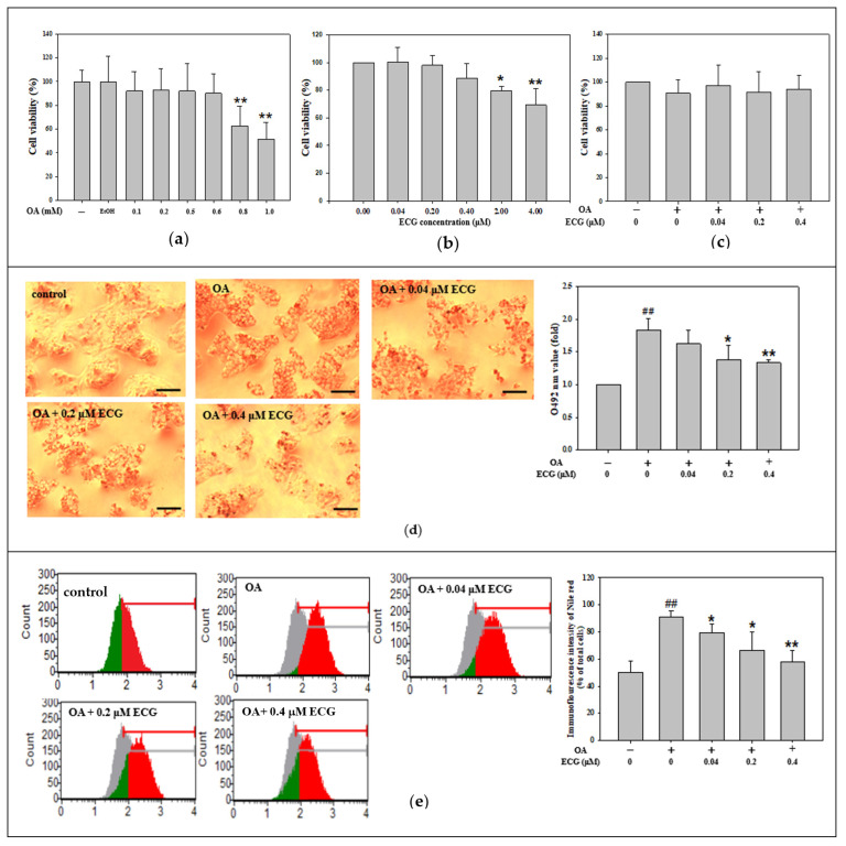
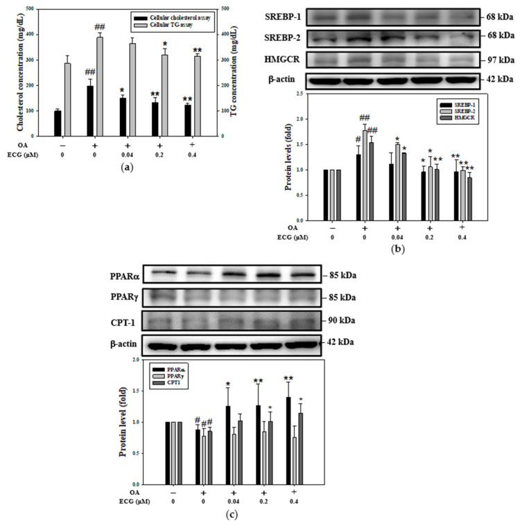
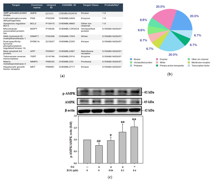
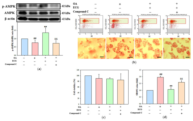
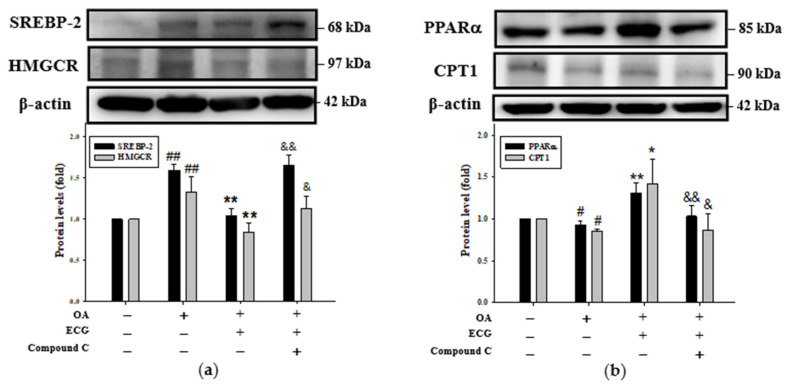
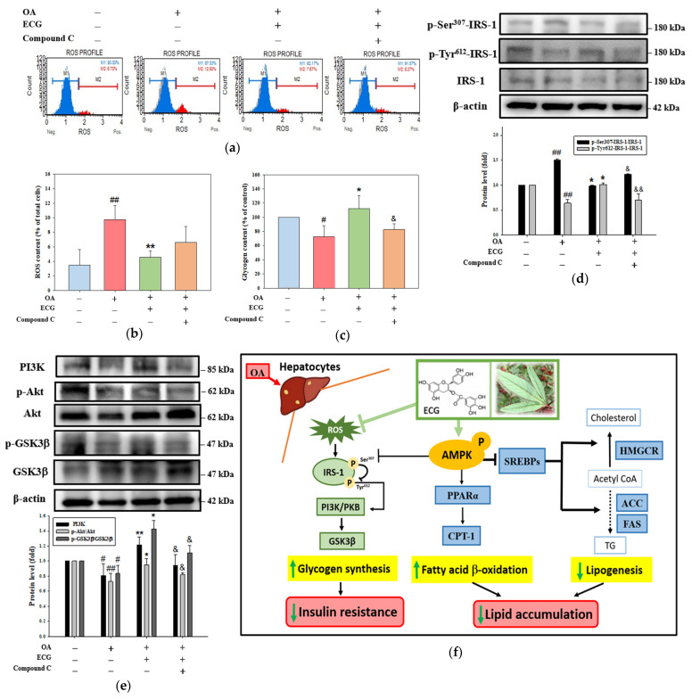
Metabolic dysfunction-associated steatotic liver disease (MASLD) begins with hepatic lipid accumulation and triggers insulin resistance. Hibiscus leaf extract exhibits antioxidant and anti-atherosclerotic activities, and is rich in (-)-epicatechin gallate (ECG). Despite ECG's well-known pharmacological activities and its total antioxidant capacity being stronger than that of other catechins, its regulatory effects on MASLD have not been fully described previously. Therefore, this study attempted to evaluate the anti-MASLD potential of ECG isolated from Hibiscus leaves on abnormal lipid and glucose metabolism in hepatocytes. First, oleic acid (OA) was used as an experimental model to induce lipid dysmetabolism in human primary hepatocytes. Treatment with ECG can significantly (p < 0.05) reduce the OA-induced cellular lipid accumulation. Nile red staining revealed, compared to the OA group, the inhibition percentages of 29, 61, and 82% at the tested doses of ECG, respectively. The beneficial effects of ECG were associated with the downregulation of SREBPs/HMGCR and upregulation of PPARα/CPT1 through targeting AMPK. Also, ECG at 0.4 µM produced a significant (p < 0.01) decrease in oxidative stress by 83%, and a marked (p < 0.05) increase in glycogen synthesis by 145% on the OA-exposed hepatocytes with insulin signaling blockade. Mechanistic assays indicated lipid and glucose metabolic homeostasis of ECG might be mediated via regulation of lipogenesis, fatty acid β-oxidation, and insulin resistance, as confirmed by an AMPK inhibitor. These results suggest ECG is a dual modulator of lipid and carbohydrate dysmetabolism in hepatocytes.

Keywords: (−)-epicatechin gallate; AMPK; Hibiscus leaves; glycogen synthesis; insulin resistance; lipid accumulation; oxidative stress.


Conflict of interest statement

The authors declare no conflicts of interest.

Figures

Figure 1
Figure 1
Effect of ECG on cell viability and lipid accumulation in the OA-challenged human primary hepatocytes. Hepatocytes were exposed to increasing concentrations of OA (0.1–1.0 mM) (a), ECG (0.04–4.0 µM) (b), or co-treated with various concentrations of ECG (0.04, 0.2, and 0.4 μM) with or without OA at 0.6 mM (c) for 24 h. EtOH served as a solvent control. Cell viability was assessed using the Muse™ Cell Analyzer. Data are shown as means ± SD (n ≥ 3) from three independent replicates. * p < 0.05, ** p < 0.01 vs. untreated group. (d) Lipid accumulation was visualized using oil red O staining and observed under a microscope at 200× magnification; scale bar, 30 μm. Red lipid droplets indicate intracellular fat. For quantification, 1 mL isopropanol was added to dissolve the stain, followed by 5× dilution in ddH2O, and absorbance was recorded at 492 nm. (e) Nile red staining followed by flow cytometric analysis was used to quantify intracellular lipid content. Data are presented as mean ± SD (n ≥ 3) from three independent replicates. ## p < 0.01 vs. control; * p < 0.05, ** p < 0.01 vs. OA group.
Figure 2
Figure 2
Effect of ECG on lipogenesis and lipolysis in the OA-treated human primary hepatocytes. Hepatocytes were exposed to different concentrations of ECG (0.04, 0.2, and 0.4 μM) with or without OA at 0.6 mM for 24 h. (a) Total cellular cholesterol (left axis) and TG (right axis) level were assayed utilizing enzymatic colorimetric assays and presented as mg/dL. The protein levels of lipogenesis-related markers (SREBP-1, SREBP-2, and HMGCR) (b) and lipolysis-associated proteins (PPARα, PPARγ, CPT1) (c) were determined by Western blotting. β-actin was used as an internal control. Results are shown as mean ± SD (n ≥ 3) from three independent replicates. # p < 0.05, ## p < 0.01 vs. control; * p < 0.05, ** p < 0.01 vs. OA group.
Figure 3
Figure 3
Involvement of AMPK in ECG’s action on the OA-treated human primary hepatocytes. * the estimated value of the most probable targets of bioactive molecules (a) The AMPK-targeting (blue marked) of ECG was analyzed utilizing the SwissTargetPrediction. (b) The distribution of predicted ECG targets across functional protein classes is shown in a pie chart. (c) Western blot analysis was performed to detect p-AMPK, total AMPK, and β-actin, an internal control, after a 24 h treatment of ECG (0.04, 0.2, and 0.4 μM) with or without OA (0.6 mM). Results are shown as mean ± SD (n ≥ 3) from three independent replicates. ## p < 0.01 vs. control; * p < 0.05, ** p < 0.01 vs. OA group.
Figure 4
Figure 4
AMPK is essential for the ECG-inhibited lipid accumulation in the OA-challenged human primary hepatocytes. Hepatocytes were pre-treated with compound C (3 µM), then co-treated with ECG (0.4 µM) and OA (0.6 mM) for 24 h. (a) The protein levels of p-AMPK and total AMPK were determined by Western blotting. β-actin was used as an internal control. The quantitative data of p-AMPK/AMPK are shown as mean ± SD (n ≥ 3) from three independent replicates. (b) Cell viability analysis (top) and oil red O staining (bottom) were performed. Images were taken at 200× magnification; scale bar, 30 μm (c) Quantitative analysis of cell viability is shown as mean ± SD (n ≥ 3) from three independent replicates. (d) Lipid staining was quantified by extracting oil red O with isopropanol and reading absorbance at 492 nm. Results are shown as mean ± SD (n ≥ 3) from three independent replicates. ## p < 0.01 vs. control; ** p < 0.01 vs. OA group; && p < 0.01 vs. OA + ECG group. (Blue column: control; Red column: OA group; Green column: OA + ECG group; Orange column: OA + ECG + Compound C group).
Figure 5
Figure 5
AMPK is essential for ECG-regulated lipogenesis and lipolysis in the OA-treated human primary hepatocytes. Hepatocytes were pre-treated with compound C (3 µM), followed by ECG (0.4 µM) and OA (0.6 mM) treatment for 24 h. Western blotting was used to assess expression of SREBP-2, HMGCR (a), PPARα, and CPT1 (b). β-actin was used as an internal control. Results are shown as mean ± SD (n ≥ 3) from three independent replicates. # p < 0.05, ## p < 0.01 vs. control; * p < 0.05, ** p < 0.01 vs. OA group; & p < 0.05, && p < 0.01 vs. OA + ECG group.
Figure 6
Figure 6
AMPK is essential for the ECG-corrected glycogen synthesis and insulin signaling in the OA-treated human primary hepatocytes. Hepatocytes were pre-treated with compound C (3 µM), followed by ECG (0.4 µM) and OA (0.6 mM) treatment for 24 h. (a) The cellular ROS were evaluated using DCFH-DA staining and flow cytometry. (b) The percentage of DCF-positive cells was calculated. (c) Cellular glycogen levels were measured using ELISA. Results are shown as mean ± SD (n ≥ 3) from three independent replicates (Blue column: control; Red column: OA group; Green column: OA + ECG group; Orange column: OA + ECG + Compound C group). Western blotting assessed levels of p-IRS-1 (Ser307 and Tyr612), IRS-1 (d), PI3K, p-PKB, PKB, p-GSK3β, and GSK3β (e). β-actin was used as an internal control. Results are shown as mean ± SD (n ≥ 3) from three independent replicates. # p < 0.05, ## p < 0.01 vs. control; * p < 0.05, ** p < 0.01 vs. OA group; & p < 0.05, && p < 0.01 vs. OA + ECG group. (f) The overview of anti-MASLD effect of ECG from Hibiscus leaves (right illustration) on the OA-induced lipid and carbohydrate dysmetabolism in hepatocytes through activating AMPK. By targeting AMPK, ECG functions against the OA’s effects involving the downregulation of hepatic lipogenesis, the upregulation of fatty acid β-oxidation (bold arrows; dashed arrows: the part to further explore), and promotion of p-Tyr-IRS-1/PKB/GSK3β axis-mediated glycogen synthesis (thin arrows), ultimately reducing lipid accumulation and insulin resistance.

Similar articles

References

    1. Friedman S.L., Neuschwander-Tetri B.A., Rinella M., Sanyal A.J. Mechanisms of NAFLD Development and therapeutic strategies. Nat. Med. 2018;24:908–922. doi: 10.1038/s41591-018-0104-9. - DOI - PMC - PubMed
    1. Rinella M.E., Lazarus J.V., Ratziu V., Francque S.M., Sanyal A.J., Kanwal F., Romero D., Abdelmalek M.F., Anstee Q.M., Arab J.P., et al. A multisociety Delphi consensus statement on new fatty liver disease nomenclature. Hepatology. 2023;78:1966–1986. doi: 10.1097/HEP.0000000000000520. - DOI - PMC - PubMed
    1. Lekakis V., Papatheodoridis G.V. Natural history of metabolic dysfunction-associated steatotic liver disease. Eur. J. Intern. Med. 2024;122:3–10. doi: 10.1016/j.ejim.2023.11.005. - DOI - PubMed
    1. Armandi A., Bugianesi E. Natural history of NASH. Liver Int. 2021;41:78–82. doi: 10.1111/liv.14910. - DOI - PMC - PubMed
    1. Wells R.G. Cellular sources of extracellular matrix in hepatic fibrosis. Clin. Liver Dis. 2008;12:759–768. doi: 10.1016/j.cld.2008.07.008. - DOI - PMC - PubMed

MeSH terms

LinkOut - more resources