Review
. 2025 Aug 6;26(15):7616.
doi: 10.3390/ijms26157616.

Glucocorticoid-Mediated Skeletal Muscle Atrophy: Molecular Mechanisms and Potential Therapeutic Targets

Affiliations
Review

Glucocorticoid-Mediated Skeletal Muscle Atrophy: Molecular Mechanisms and Potential Therapeutic Targets

Uttapol Permpoon et al. Int J Mol Sci. .

Abstract

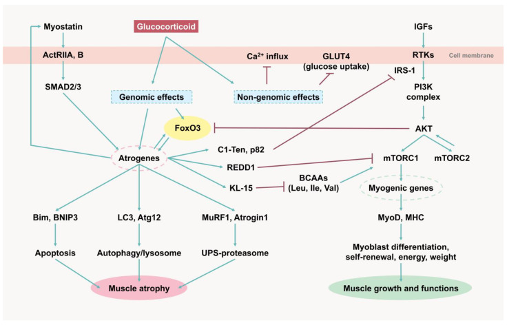
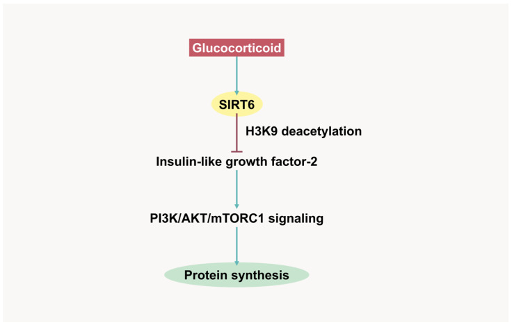
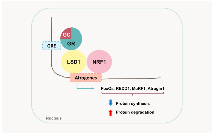
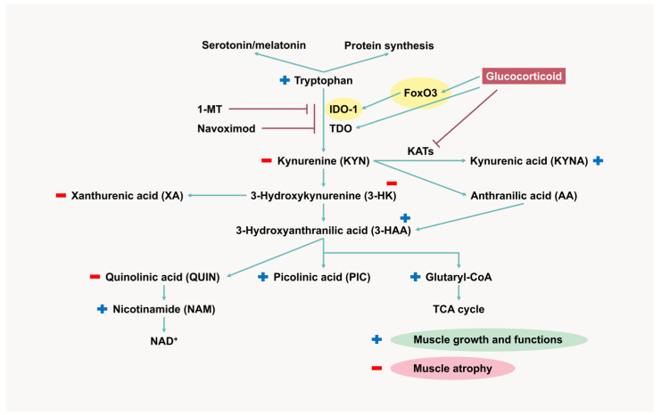
Skeletal muscle atrophy is a critical health issue affecting the quality of life of elderly individuals and patients with chronic diseases. These conditions induce dysregulation of glucocorticoid (GC) secretion. GCs play a critical role in maintaining homeostasis in the stress response and glucose metabolism. However, prolonged exposure to GC is directly linked to muscle atrophy, which is characterized by a reduction in muscle size and weight, particularly affecting fast-twitch muscle fibers. The GC-activated glucocorticoid receptor (GR) decreases protein synthesis and facilitates protein breakdown. Numerous antagonists have been developed to mitigate GC-induced muscle atrophy, including 11β-HSD1 inhibitors and myostatin and activin receptor blockers. However, the clinical trial results have fallen short of the expected efficacy. Recently, several emerging pathways and targets have been identified. For instance, GC-induced sirtuin 6 isoform (SIRT6) expression suppresses AKT/mTORC1 signaling. Lysine-specific demethylase 1 (LSD1) cooperates with the GR for the transcription of atrogenes. The kynurenine pathway and indoleamine 2,3-dioxygenase 1 (IDO-1) also play crucial roles in protein synthesis and energy production in skeletal muscle. Therefore, a deeper understanding of the complexities of GR transactivation and transrepression will provide new strategies for the discovery of novel drugs to overcome the detrimental effects of GCs on muscle tissues.

Keywords: IDO-1; LSD1; SIRT6; atrogenes; glucocorticoids; muscle atrophy.


Conflict of interest statement

The authors report no conflicts of interest.

Figures

Figure 1
Figure 1
Molecular mechanisms of glucocorticoid-induced muscle atrophy. ActRIIA, B, activin type II receptor A and B; GLUT4, glucose transporter 4; IGFs, insulin-like growth factors; RTKs, receptor tyrosine kinases; IRS-1, insulin receptor substrate-1; PI3K, phosphatidylinositol 3-kinase; AKT, protein kinase B (PKB); mTORC, mammalian target of rapamycin complex; MyoD, myoblast determination protein 1; MHC, myosin heavy chain; FoxO3, Forkhead Box O 3; REDD1, regulated in development and DNA damage 1; KLF-15, Kruppel-like factor 15; BCAAs, branched-chain amino acids; MuRF1, Muscle RING Finger 1; Atrogin1, Muscle Atrophy F-box (MAFbx).
Figure 2
Figure 2
Glucocorticoid-induced SIRT6 expression in muscle protein synthesis suppression. SIRT6, Sirtuin 6; H3K9, histone 3 lysine 9; PI3K, phosphatidylinositol 3-kinase; AKT, protein kinase B (PKB); mTORC1, mammalian target of rapamycin complex 1.
Figure 3
Figure 3
The cooperation of GR, LSD1, and NRF1 in the activation of atrogenes and enhancement of protein degradation. GRE, glucocorticoid response element; GC, glucocorticoid; GR, glucocorticoid receptor; LSD1, lysine-specific demethylase 1; NRF1, nuclear respiratory factor 1; FoxOs, Forkhead Box O members; REDD1, regulated in development and DNA damage 1; MuRF1, Muscle RING Finger 1; Atrogin1, Muscle Atrophy F-box (MAFbx).
Figure 4
Figure 4
Role of the kynurenine pathway in muscle biology and effects of glucocorticoid on kynurenine metabolites. FoxO3, Forkhead Box O 3; IDO-1, indoleamine 2,3-dioxygenase 1; TDO, tryptophan 2,3-dioxygenase; KATs, KYN aminotransferases; NAD, nicotinamide adenine dinucleotide; TCA, tricarboxylic acid cycle; 1-MT, 1-methyl-tryptophan.

Similar articles

References

    1. Yin L., Li N., Jia W., Wang N., Liang M., Yang X., Du G. Skeletal muscle atrophy: From mechanisms to treatments. Pharmacol. Res. 2021;172:105807. doi: 10.1016/j.phrs.2021.105807. - DOI - PubMed
    1. Ahmad S.S., Chun H.J., Ahmad K., Shaikh S., Lim J.H., Ali S., Han S.S., Hur S.J., Sohn J.H., Lee E.J., et al. The roles of growth factors and hormones in the regulation of muscle satellite cells for cultured meat production. J. Anim. Sci. Technol. 2023;65:16–31. doi: 10.5187/jast.2022.e114. - DOI - PMC - PubMed
    1. Yoshida T., Delafontaine P. Mechanisms of IGF-1-Mediated Regulation of Skeletal Muscle Hypertrophy and Atrophy. Cells. 2020;9:1970. doi: 10.3390/cells9091970. - DOI - PMC - PubMed
    1. Lee J.H., Jun H.S. Role of myokines in regulating skeletal muscle mass and function. Front. Physiol. 2019;10:42. doi: 10.3389/fphys.2019.00042. - DOI - PMC - PubMed
    1. Zhang H., Qi G., Wang K., Yang J., Shen Y., Yang X., Chen X., Yao X., Gu X., Qi L., et al. Oxidative stress: Roles in skeletal muscle atrophy. Biochem. Pharmacol. 2023;214:115664. doi: 10.1016/j.bcp.2023.115664. - DOI - PubMed

LinkOut - more resources