Review
. 2025 Aug 7;26(15):7642.
doi: 10.3390/ijms26157642.

Molecular Crosstalk Between RUNX2 and HIF-1α in Osteosarcoma: Implications for Angiogenesis, Metastasis, and Therapy Resistance

Affiliations
Review

Molecular Crosstalk Between RUNX2 and HIF-1α in Osteosarcoma: Implications for Angiogenesis, Metastasis, and Therapy Resistance

Anuja Gajanan Magar et al. Int J Mol Sci. .

Abstract

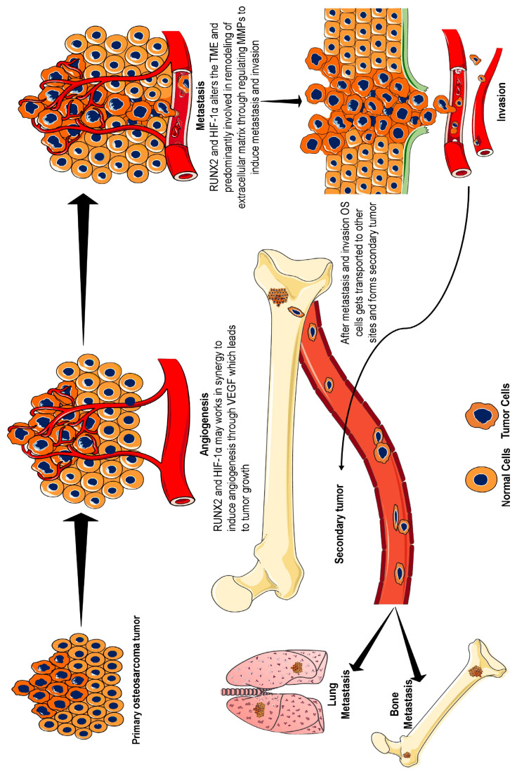
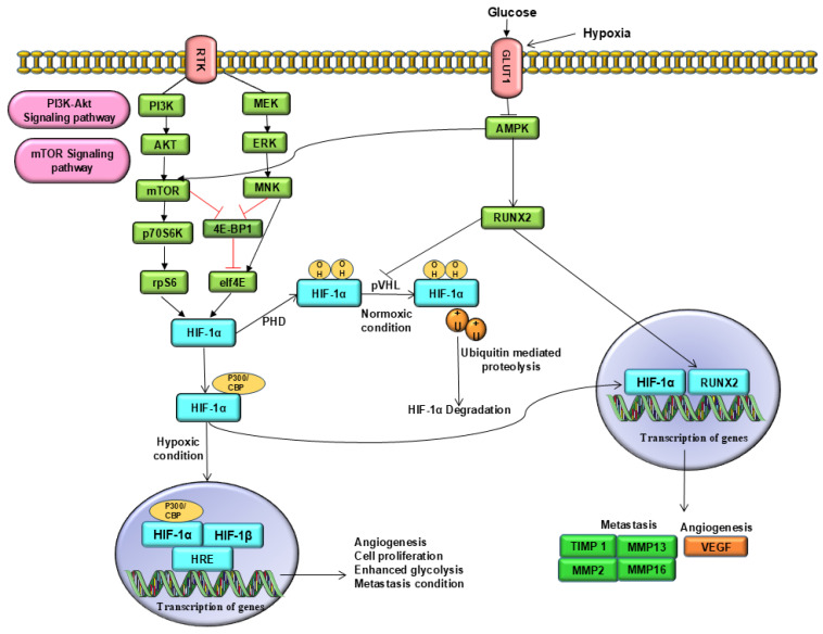
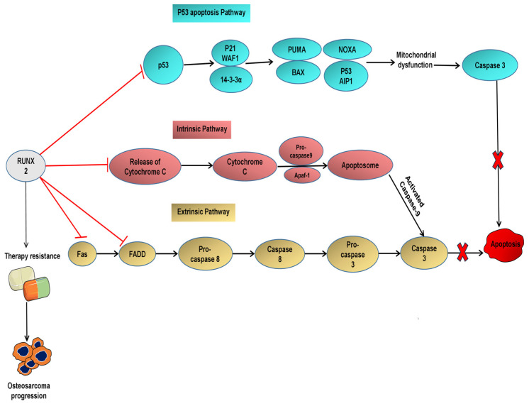
Runt-related transcription factor-2 (RUNX2) is an integral player in osteogenesis and is highly expressed in osteosarcoma. Emerging evidence suggests that aberrant RUNX2 expression is a key factor in osteosarcoma oncogenesis. Patients with advanced stages of osteosarcoma overexpressing RUNX2 are more likely to have high tumour grades, metastasis, and lower overall or progression-free survival rates. Thus, RUNX2 is considered a potential candidate for targeted therapy of osteosarcoma. Hypoxia-inducible factor-1α (HIF-1α) is a key transcription factor involved in the regulation of cellular reprogramming in response to hypoxia. Overexpression of HIF-1α decreases overall survival, disease-free survival, and chemotherapy response and promotes tumour stage and metastasis. Hence, our review focused on highlighting the intricate network between RUNX2 and HIF-1α, which support each other or may work synergistically to develop resistance to therapy and osteosarcoma progression. An in-depth understanding of these two important tumour progression markers is required. Therefore, this review focuses on the role of RUNX2 and HIF-1α in the alteration of the tumour microenvironment, which further promotes angiogenesis, metastasis, and resistance to therapy in osteosarcoma.

Keywords: HIF-1α; RUNX2; angiogenesis; metastasis; osteosarcoma; therapy resistance.


Conflict of interest statement

The authors declare no conflicts of interest.

Figures

Figure 1
Figure 1
Development and progression of osteosarcoma. Under hypoxic conditions, HIF-1α is induced and creates a tumour-friendly environment through aberrant angiogenesis via VEGF signalling. RUNX2 also plays an active role, or it may be that both of them work in synergy to stabilize VEGF, an angiogenesis precursor. Furthermore, HIF-1α and RUNX2 alter the tumour microenvironment through enhanced glycolysis and induce MMP expression, leading to extracellular matrix remodelling. MMP causes metastasis and invasion, which helps cancer cells to transport to other bone and lung sites. In osteosarcoma, the primary site of metastasis is the lung, followed by the other bones. Therefore, the overexpression of RUNX2 and HIF-1α can be considered a biomarker for osteosarcoma because of their roles in the stimulation of angiogenesis, metastasis, and invasion. Abbreviations: HIF-1α, hypoxia-inducible factor-1α; RUNX2, runt-related transcription factor-2; VEGF, vascular endothelial growth factor; MMP, matrix metalloproteinase.
Figure 2
Figure 2
Molecular crosstalk between RUNX2 and HIF-1α in osteosarcoma. Under normoxic conditions, HIF-1α is hydroxylated by PHDs. PHDs hydroxylate proline residues and provide a binding site for pVHL, a tumour suppressor protein. This tumour suppressor gene leads to polyubiquitination and proteasomal degradation of HIF-1α. This hydroxylation impedes the binding of HIF-1α to the transcriptional coactivators p300 and CBP. In contrast, under hypoxic conditions, PHDs are unable to hydroxylate HIF-1α. Therefore, HIF-1α translocates to the nucleus and dimerises with HIF-β subunit. This complex activates the HIF pathway and promotes the transcription of genes that enhance glucose metabolism, angiogenesis, metastasis, and invasiveness. In addition, RUNX2 competes with the E3 ubiquitin ligase pVHL to prevent the degradation of HIF-1α. This stabilized expression of HIF-1α further enhances glycolysis via overexpression of GLUT1 receptors and enhances glucose transport. This will aid in the metabolic shift towards aerobic glycolysis and support rapid tumour growth. Additionally, the activity of AMPK, a negative regulator of mTOR, and RUNX2 is suppressed under hypoxic conditions, leading to the stabilization of the mTOR pathway responsible for cell proliferation. Downregulation of AMPK prevents degradation and helps in RUNX2 stabilization. This synergistic interaction between RUNX2 and HIF-1α induces VEGF expression and promotes angiogenesis, metastasis, and invasion. Abbreviations: AMPK, adenosine monophosphate-activated protein kinase; CBP: Core binding protein; GLUT1, glucose transporter 1; HIF-1α, hypoxia inducible factor-1α; mTOR, mammalian targets of rapamycin; PHDs, prolyl hydroxylase domain; PI3K/AKT: Phosphatidylinositol 3-kinase/Protein kinase B; RUNX2, runt-related transcription factor 2; pVHL, von Hippel-Lindau protein; VEGF, vascular endothelial growth factor.
Figure 3
Figure 3
The role of RUNX2 in the development of therapy resistance RUNX2 prevents osteosarcoma cells from apoptosis by suppressing the activity of caspases and p-53 targeted genes. For example, RUNX2 downregulates the activation of p53-targeted genes including p21, WAF1, PUMA, and NOXA, which are responsible for apoptosis. In addition, overexpression of RUNX2 prevents the release of the extrinsic ligand Fas, adaptor protein FADD, and intrinsic apoptotic marker cytochrome C. It prevents the activation of caspase-3, caspase-8 and caspase-9. Thus, RUNX2 inhibits the intrinsic and extrinsic apoptotic pathways and protects osteosarcoma cells from apoptosis after chemotherapy. This implies that the regulation of RUNX2 in osteosarcoma will help enhance chemosensitivity and reactivate the apoptosis pathway. Abbreviations: Apaf-1: Apoptotic Protease Activating Factor 1; Bax, BCL-2-associated X protein; Fas, FS-7-associated surface antigen; FADD, Fas-associated death domain protein; NOXA, Phorbol-12-myristate-13-acetate-induced protein 1; PUMA: p53 upregulated modulator of apoptosis; RUNX2: runt-related transcription factor 2; WAF1: Wild-type Allele Frequency 1.

Similar articles

References

    1. Idoate M.Á., Aquerreta J.D., Lamo-Espinosa J.M., San-Julian M. A Reassessment of the Barrier Effect of the Physis against Metaphyseal Osteosarcoma: A Comprehensive Pathological Study with Its Radiological and Clinical Follow-Up Correlations. Diagnostics. 2022;12:450. doi: 10.3390/diagnostics12020450. - DOI - PMC - PubMed
    1. Prabowo Y., Primaputra M.R.A., Kodrat E. Reconstruction of osteosarcoma of the proximal tibia using bone on polyethylene hemiarthroplasty knee joint system: A case report. Int. J. Surg. Case Rep. 2020;72:188–196. doi: 10.1016/j.ijscr.2020.05.013. - DOI - PMC - PubMed
    1. Chen C., Xie L., Ren T., Huang Y., Xu J., Guo W. Immunotherapy for osteosarcoma: Fundamental mechanism, rationale, and recent breakthroughs. Cancer Lett. 2021;500:1–10. doi: 10.1016/j.canlet.2020.12.024. - DOI - PubMed
    1. Eaton B.R., Schwarz R., Vatner R., Yeh B., Claude L., Indelicato D.J., Laack N. Osteosarcoma. Pediatr. Blood Cancer. 2021;68:e28352. doi: 10.1002/pbc.28352. - DOI - PubMed
    1. Tuck S.P., Walker J. Adult Paget’s disease of bone. Clin. Med. 2020;20:568–571. doi: 10.7861/clinmed.20.6.page. - DOI - PMC - PubMed

LinkOut - more resources