. 2025 Aug 7;26(15):7659.
doi: 10.3390/ijms26157659.

Immunohistochemical Analysis of Placental Tissue of Women Infected with SARS-CoV-2 During Pregnancy-A Prospective Clinical Study

Affiliations

Immunohistochemical Analysis of Placental Tissue of Women Infected with SARS-CoV-2 During Pregnancy-A Prospective Clinical Study

Marija Bicanin Ilic et al. Int J Mol Sci. .

Abstract

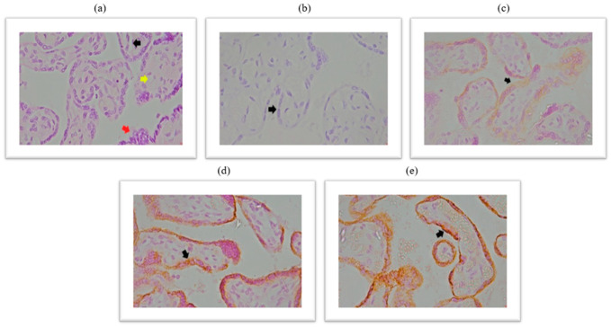
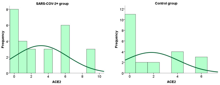
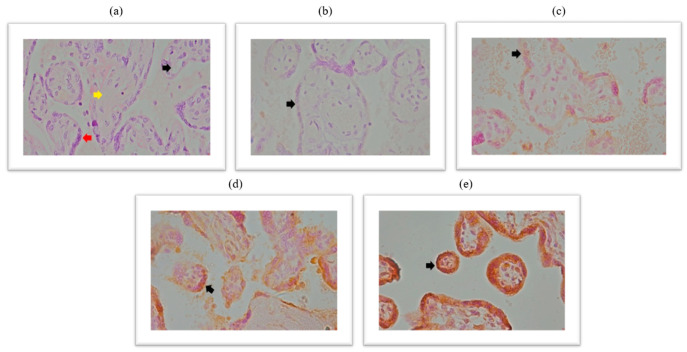
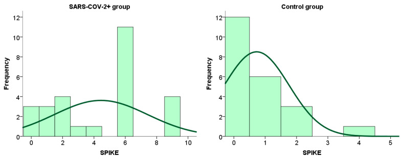
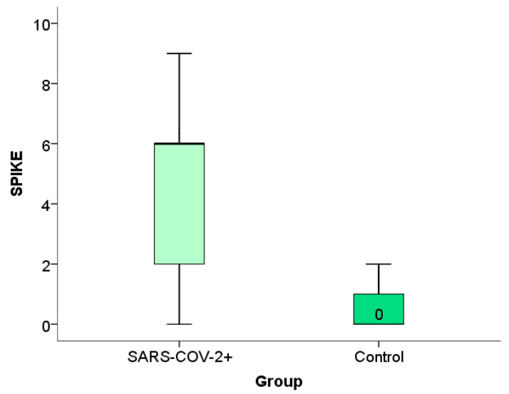
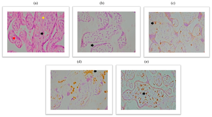
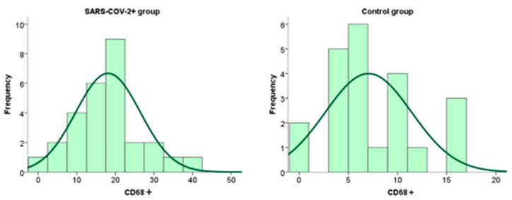
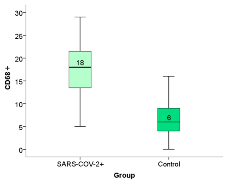
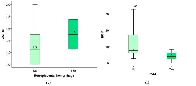
SARS-CoV-2 has an affinity for binding to the human Angiotensin-converting enzyme 2 (ACE2) receptor through cleavage and conformational changes at the S1-S2 boundary and the receptor binding domain of the spike protein, which is also the most variable part of SARS-CoV-2. This study aimed to investigate the expression of Angiotensin-converting enzyme 2 (ACE2), spike protein, and CD68+ markers in placental tissue to demonstrate a possible correlation with the level of systemic oxidative stress biomarkers in patients who were infected with SARS-CoV-2 during pregnancy. A prospective clinical cohort study was designed to investigate the presence of CD68+ macrophages, ACE2, and spike proteins in placental tissue using immunohistochemical methods and to compare these results with oxidative stress from our previous study. Spike and CD68+ macrophages' immunoreactivity were more pronounced in the placental tissue of patients from the SARS-CoV-2 group. Placental tissue spike protein and CD68+ immunoreactivity correlate with maternal and fetal Thiobarbituric Acid Reactive (TBARS) levels. This study has confirmed that spike protein expression in placental tissue is associated with the newborn's stay in intensive neonatal care. Therefore, immunoreactivity analysis for the Spike antigen is important in detecting newborns at risk of early neonatal complications.

Keywords: ACE2 receptor; CD68+ leucocytes; SARS-CoV-2; placental inflammation; spike.


Conflict of interest statement

The authors declare no conflicts of interest.

Figures

Figure 1
Figure 1
(a) Chorionic villi with edematous stroma and dilatated blood vessels, HE stained, magnification 400×; (b) trophoblast cells do not show membrane and cytoplasmic immunopositivity on ACE2 antibody-negative control, magnification 400×; (c) trophoblast cells show membrane and cytoplasmic immunopositivity on ACE2 antibody of light intensity (light yellow color), magnification 400×; (d) trophoblast cells show membrane and cytoplasmic immunopositivity on ACE2 antibody of moderate intensity (light brown color), magnification 400×; (e) trophoblast cells show membrane and cytoplasmic immunopositivity on ACE2 antibody of pronounced intensity (dark brown color), magnification of 400×. Black arrow represents dilated blood vessels; yellow arrow represents edematous stroma; red arrow represents cytotrophoblast cells.
Figure 2
Figure 2
Frequency of ACE2 expression on placental tissue cells in patients infected with SARS-CoV-2 during pregnancy and in the control group.
Figure 3
Figure 3
(a) Chorionic villi with edematous stroma and dilatated blood vessels, HE stained, magnification 400×; (b) trophoblast cells do not show immunoreactivity to SARS-CoV-2 spike antibody-negative control, magnification 400×; (c) trophoblast cells exhibit membrane and cytoplasmic immunopositivity of light intensity to SARS-CoV-2 spike antibody (light yellow color), magnification 400×; (d) trophoblast cells show membrane and cytoplasmic immunopositivity of moderate intensity to SARS-CoV-2 spike antibody (light brown color), 400× magnification; (e) trophoblast cells show membrane and cytoplasmic immunopositivity of pronounced intensity on SARS-CoV-2 spike antibody (dark brown color), magnification 400×. Black arrow represents dilated blood vessels; yellow arrow represents edematous stroma; red arrow represents cytotrophoblast cells.
Figure 4
Figure 4
Frequency of spike protein expression within placental tissue in patients infected with SARS-CoV-2 during pregnancy and in the control group.
Figure 5
Figure 5
Distribution of frequency of spike protein expression within placental tissue in patients infected with SARS-CoV-2 during pregnancy and in the control group.
Figure 6
Figure 6
(a): The 1a chorionic villi with edematous stroma and dilatated blood vessels, HE stained, magnification 400×; (b) negative control, magnification 400×; (b) rare CD68+ macrophages in stroma and intervillous spaces, magnification 400×; (c) numerous CD68+ macrophages are present in the intervillous spaces, covering more than 5% of the intervillous space-chronic histiocytic intervillitis, magnification 400×; (d) chronic villous histiocytosis, magnification 400×. The black arrow represents dilated blood vessels; yellow arrow represents edematous stroma; red arrow represents cytotrophoblast cells. Black arrow (d,e)—numerous Hoffbauer cells–fetal macrophages located in stroma of villi.
Figure 7
Figure 7
Frequency of expression of CD68+ macrophages within placental tissue in patients infected with SARS-CoV-2 during pregnancy and in the control group.
Figure 8
Figure 8
Distribution of the degree of expression of CD68+ macrophage immunoreactivity within placental tissue in patients infected with SARS-CoV-2 during pregnancy and in the control group.
Figure 9
Figure 9
(a) Correlation between placental weight and week of gestation in which the infection occurred (p = 0.17); (b) correlation between the ratio of placental weight to child weight and week of gestation in which the infection occurred (p = 0.33).
Figure 10
Figure 10
Correlation of the spike protein expression within placental tissue with the stay of the newborn in neonatal intensive care (a) and with the expression of CD68+ macrophages (b).
Figure 11
Figure 11
Correlation curve between ACE2 with O2 anion expression in maternal serum (a), CD68+ macrophage expression in placental tissue with maternal serum TBARS levels (b), CD68+ macrophage expression in placental tissue with umbilical cord blood TBARS level (c).
Figure 12
Figure 12
(a) The level of TBARS of the newborn concerning the presence of avascular villi (p = 0.05); (b) the association between TBARS of the newborn and obliteration of blood vessels (p = 0.04); (c) the association between maternal TBARS and vascular ecstasy (p = 0.03); (d) the association between maternal TBARS and placental infarction (p = 0.03); (e) the association between the level of maternal catalase and retroplacental hemorrhage (p = 0.03); (f) the association between the newborn NO of the fetus with the presence of FVM (p = 0.00).
Figure 12
Figure 12
(a) The level of TBARS of the newborn concerning the presence of avascular villi (p = 0.05); (b) the association between TBARS of the newborn and obliteration of blood vessels (p = 0.04); (c) the association between maternal TBARS and vascular ecstasy (p = 0.03); (d) the association between maternal TBARS and placental infarction (p = 0.03); (e) the association between the level of maternal catalase and retroplacental hemorrhage (p = 0.03); (f) the association between the newborn NO of the fetus with the presence of FVM (p = 0.00).

Similar articles

References

    1. Macáková K., Pšenková P., Šupčíková N., Vlková B., Celec P., Záhumenský J. Effect of SARS-CoV-2 Infection and COVID-19 Vaccination on Oxidative Status of Human Placenta: A Preliminary Study. Antioxidants. 2023;12:1403. doi: 10.3390/antiox12071403. - DOI - PMC - PubMed
    1. China CDC Weekly Protocol for Prevention and Control of COVID-19 (Edition 6) China CDC Wkly. 2020;2:321–326. doi: 10.46234/ccdcw2020.082. - DOI - PMC - PubMed
    1. Asselah T., Durantel D., Pasmant E., Lau G., Schinazi R.F. COVID-19: Discovery, Diagnostics and Drug Development. J. Hepatol. 2021;74:168–184. doi: 10.1016/j.jhep.2020.09.031. - DOI - PMC - PubMed
    1. Zang Y., Gu L., Zhang Y., Wang Y., Xue F. Identification of Key Genes and Pathways in Uterine Leiomyosarcoma through Bioinformatics Analysis. Oncol. Lett. 2018;15:9361–9368. doi: 10.3892/ol.2018.8503. - DOI - PMC - PubMed
    1. Bicanin Ilic M., Nikolic Turnic T., Ilic I., Nikolov A., Mujkovic S., Rakic D., Jovic N., Arsenijevic N., Mitrovic S., Spasojevic M., et al. SARS-CoV-2 Infection and Its Association with Maternal and Fetal Redox Status and Outcomes: A Prospective Clinical Study. J. Clin. Med. 2025;14:1555. doi: 10.3390/jcm14051555. - DOI - PMC - PubMed

LinkOut - more resources