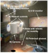
Can 3D Exoscopy-Assisted Surgery Replace the Traditional Endoscopy in Septoplasty? Analysis of Our Two-Year Experience
- PMID: 40806901
- PMCID: PMC12347903
- DOI: 10.3390/jcm14155279
Can 3D Exoscopy-Assisted Surgery Replace the Traditional Endoscopy in Septoplasty? Analysis of Our Two-Year Experience
Abstract
Background/Objectives: Septoplasty is a commonly performed surgical procedure aimed at correcting nasal septal deviations, to improve nasal airflow and respiratory function. Traditional approaches to septal correction rely on either direct visualization or endoscopic guidance. Recently, a novel technology known as exoscopy has been introduced into surgical practice. Exoscopy is an "advanced magnification system" that provides an enlarged, three-dimensional view of the operating field. In this article, we present our experience with exoscope-assisted septoplasty, developed over the last two years, and compare it with our extensive experience using the endoscopic approach. Methods: Our case series includes 26 patients, predominantly males and young adults, who underwent exoscope-assisted septoplasty. We discuss the primary advantages of this technique and, most importantly, provide an analysis of its learning curve. The cohort of patients treated using the exoscopic approach was compared with a control group of 26 patients who underwent endoscope-guided septoplasty, randomly selected from our broader clinical database. Finally, we present a representative surgical case that details all phases of the exoscope-assisted procedure. Results: Our surgical experience has demonstrated that exoscopy is a safe and effective tool for performing septoplasty. Moreover, the learning curve associated with this technique exhibits a rapid and progressive improvement. Notably, exoscopy provides a substantial educational benefit for trainees and medical students, as it enables them to share the same visual perspective as the lead surgeon. Conclusions: Although further studies are required to validate this approach, we believe that exoscopy represents a promising advancement for a wide range of head and neck procedures, and certainly for septoplasty.
Keywords: 3D surgery; exoscopy; magnification; new technologies; rhinoseptoplasty.
Conflict of interest statement
The authors declare no conflict of interest.
Figures


Similar articles
-
Prescription of Controlled Substances: Benefits and Risks.2025 Jul 6. In: StatPearls [Internet]. Treasure Island (FL): StatPearls Publishing; 2025 Jan–. 2025 Jul 6. In: StatPearls [Internet]. Treasure Island (FL): StatPearls Publishing; 2025 Jan–. PMID: 30726003 Free Books & Documents.
-
New 3-D Technologies in Salivary Gland Surgery: The Exoscope-Assisted Surgery for Treatment of Benign Tumors of Parotid Gland.Indian J Otolaryngol Head Neck Surg. 2024 Oct;76(5):4506-4515. doi: 10.1007/s12070-024-04898-z. Epub 2024 Jul 20. Indian J Otolaryngol Head Neck Surg. 2024. PMID: 39376411
-
Multilevel-Single Stage-Functional Rhinoplasty & BRP (Barb Reposition Palatoplasty) in Surgical Management of Primary Snorers, UARS and Mild OSA.Indian J Otolaryngol Head Neck Surg. 2024 Dec;76(6):5672-5681. doi: 10.1007/s12070-024-05059-y. Epub 2024 Oct 8. Indian J Otolaryngol Head Neck Surg. 2024. PMID: 39559159
-
Systemic pharmacological treatments for chronic plaque psoriasis: a network meta-analysis.Cochrane Database Syst Rev. 2021 Apr 19;4(4):CD011535. doi: 10.1002/14651858.CD011535.pub4. Cochrane Database Syst Rev. 2021. Update in: Cochrane Database Syst Rev. 2022 May 23;5:CD011535. doi: 10.1002/14651858.CD011535.pub5. PMID: 33871055 Free PMC article. Updated.
-
Systemic pharmacological treatments for chronic plaque psoriasis: a network meta-analysis.Cochrane Database Syst Rev. 2020 Jan 9;1(1):CD011535. doi: 10.1002/14651858.CD011535.pub3. Cochrane Database Syst Rev. 2020. Update in: Cochrane Database Syst Rev. 2021 Apr 19;4:CD011535. doi: 10.1002/14651858.CD011535.pub4. PMID: 31917873 Free PMC article. Updated.
References
-
- Peynègre R., Bossard B., Koskas G., Borsik M., Bouaziz A., Gilain L. La chirurgie endoscopique des cornets. Etude préliminaire [Endoscopic surgery of the turbinates. Preliminary results] Ann. Otolaryngol. Chir. Cervicofac. 1989;106:537–540. - PubMed
-
- Bollobas B. Arcüreg punctiós troicar és endoscop [Trocar puncture and endoscopy of the maxillary sinus] Orv. Hetil. 1953;94:1399–1400. - PubMed
LinkOut - more resources
Full Text Sources

