. 2025 Jul 25;14(15):2610.
doi: 10.3390/foods14152610.

Occurrence of Aspergillus and Penicillium Species, Accumulation of Fungal Secondary Metabolites, and qPCR Detection of Potential Aflatoxigenic Aspergillus Species in Chickpea (Cicer arietinum L.) Seeds from Different Farming Systems

Affiliations

Occurrence of Aspergillus and Penicillium Species, Accumulation of Fungal Secondary Metabolites, and qPCR Detection of Potential Aflatoxigenic Aspergillus Species in Chickpea (Cicer arietinum L.) Seeds from Different Farming Systems

Mara Quaglia et al. Foods. .

Abstract

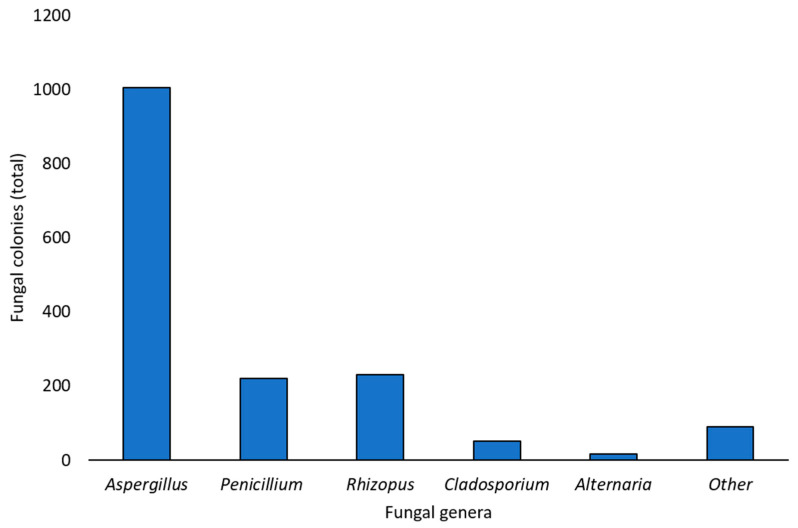
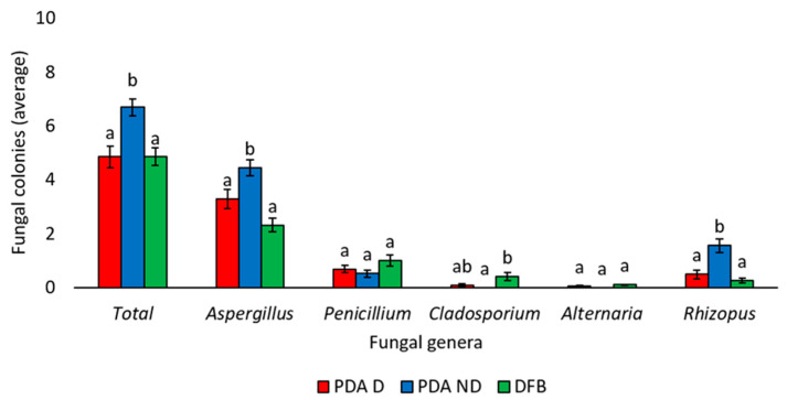
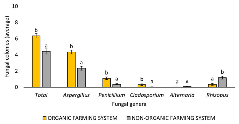
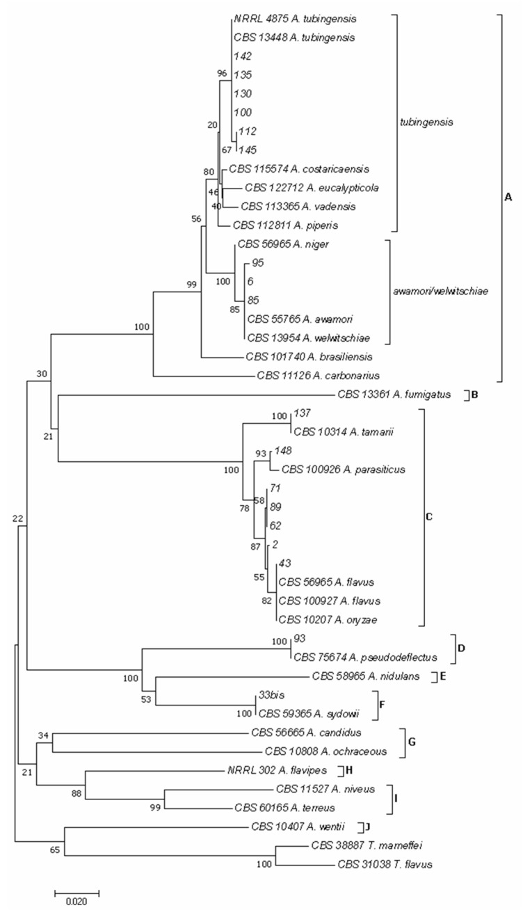
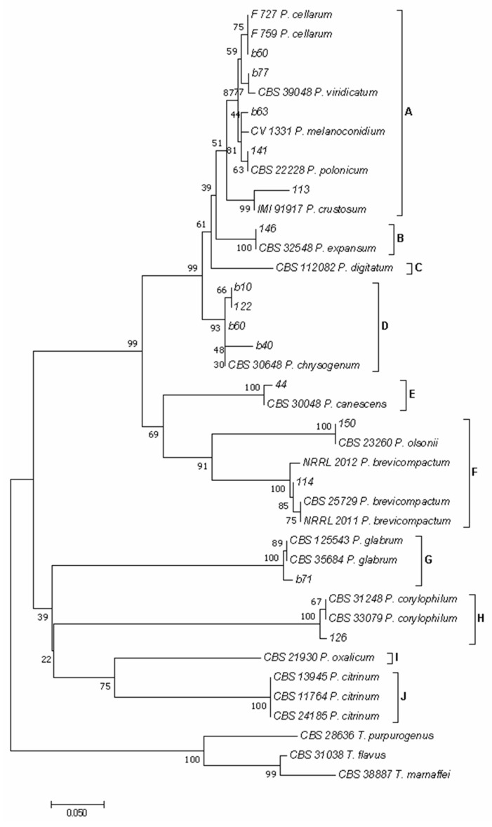
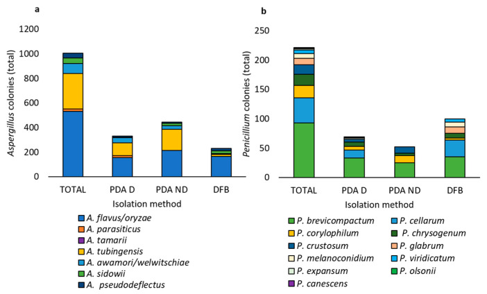
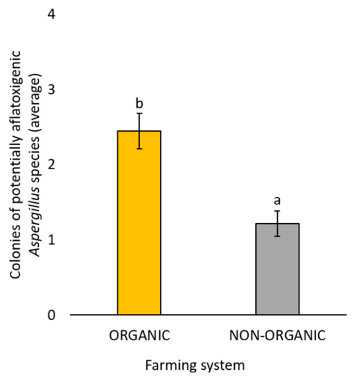
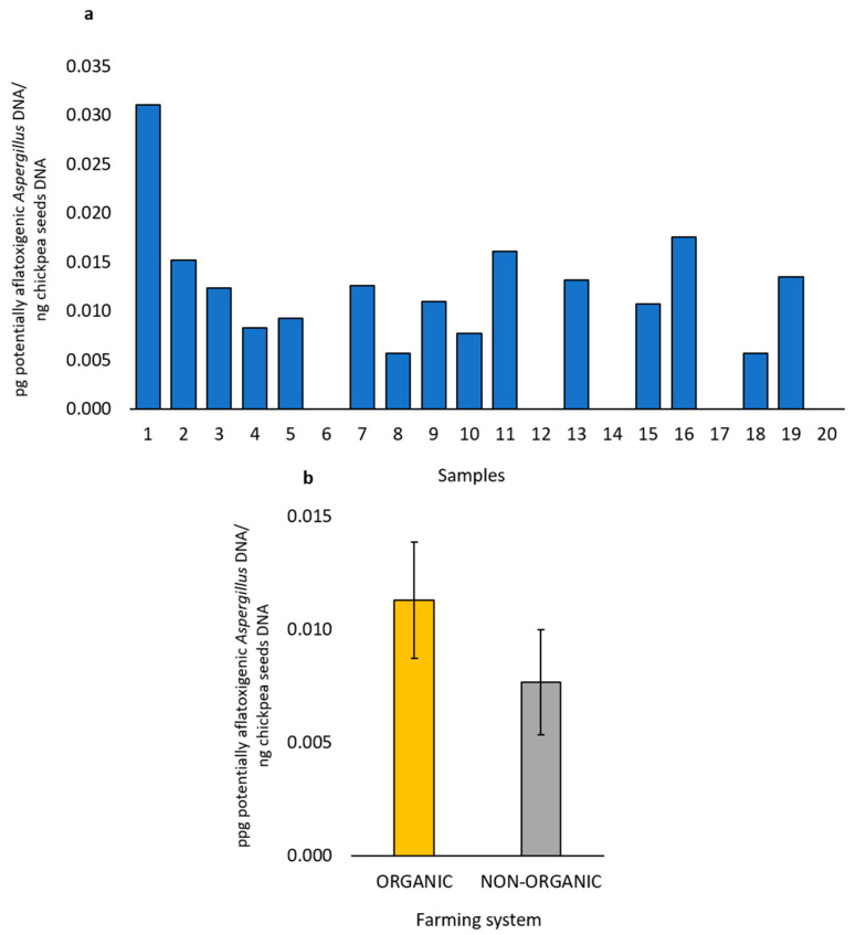
The European chickpea market raises concerns about health risks for consumers due to contamination by mycotoxins. Contamination levels can vary depending on the farming system, and rapid and reliable screening tools are desirable. In this study, marketed chickpea seed samples from organic and non-organic farming systems were analyzed for fungal and mycotoxin contamination. Aspergillus and Penicillium were the most frequently identified mycotoxigenic genera. Significant differences in fungal detection were observed among the three isolation methods used, whose combined application is proposed to enhance detection efficiency. The number of Aspergillus and Penicillium colonies was significantly higher in the organic samples. Molecular analysis identified different species within each genus, including several not previously reported in chickpea, as well as potentially aflatoxigenic species such as A. flavus/oryzae and A. parasiticus. LC-MS/MS analysis revealed aflatoxin production only by A. parasiticus, which was present in low amounts. However, the presence of potentially aflatoxigenic Aspergillus species suggests that chickpeas should be monitored to detect their safety and subsequently protect consumer health. A qPCR protocol targeting the omt-1 gene, involved in aflatoxin biosynthesis, proved to be a promising rapid tool for detecting potentially aflatoxigenic Aspergillus species.

Keywords: aflatoxins; mycobiota; mycotoxigenic fungi; mycotoxins; organic farming system; post-harvest; pulses.


Conflict of interest statement

The authors declare no conflicts of interest.

Figures

Figure 1
Figure 1
Total number of fungal colonies developed from the total marketed chickpea seed samples with all the isolation techniques used in this survey (Potato Dextrose Agar with surface disinfection, Potato Dextrose Agar without surface disinfection, deep-freezing blotter) and identified as belonging to different genera or not assigned to any genus (other) based on the observation of morphological features.
Figure 2
Figure 2
Average number of total fungal colonies or fungal colonies belonging to the Aspergillus, Penicillium, Cladosporium, Alternaria, and Rhizopus genera developed from marketed chickpea seed samples using three different isolation methods: Potato Dextrose Agar with surface disinfection (PDA D), Potato Dextrose Agar without surface disinfection (PDA ND), and the deep-freezing blotter (DFB). The genus of each fungal colony was morphologically identified. Each column represents the average (±standard error) of the 20 analyzed samples. Within each category (Total, Aspergillus, Penicillium, Cladosporium, Alternaria, Rhizopus), different letters indicate significant differences (p < 0.05, Tukey’s HSD test) among the three different isolation methods (PDA D, PDA ND, or DFB).
Figure 3
Figure 3
Average number of total fungal colonies or fungal colonies belonging to the Aspergillus, Penicillium, Cladosporium, Alternaria, and Rhizopus genera developed following the use of all three different isolation methods (Potato Dextrose Agar with surface disinfection, Potato Dextrose Agar without surface disinfection, and deep-freezing blotter) from marketed chickpea seed samples obtained with organic or non-organic farming systems. The genus of each fungal colony was morphologically identified. Each column represents the average (±standard error) of the 10 analyzed samples per each farming system. Different letters indicate significant differences (p < 0.05, Tukey’s HSD test) between the two different farming systems within each category (Total, Aspergillus, Penicillium, Cladosporium, Alternaria, Rhizopus).
Figure 4
Figure 4
Phylogeny of Aspergillus isolates obtained from the marketed chickpea seed samples surveyed in this study. The evolutionary history of Aspergillus isolates was inferred using the Neighbor-Joining method [39] and a combined dataset of BenA and CaM gene sequences. The optimal tree with the sum of branch length = 1.35228509 is shown. The percentage of replicate trees in which the associated taxa clustered together in the bootstrap test (1000 replicates) are shown next to the branches [40]. The tree is drawn to scale, with branch lengths in the same units as those of the evolutionary distances used to infer the phylogenetic tree. The evolutionary distances were computed using the Maximum Composite Likelihood method [41] and are in the units of the number of base substitutions per site. The analysis involved 46 nucleotide sequences. All positions containing gaps and missing data were eliminated. There was a total of 451 positions in the final dataset. Evolutionary analyses were conducted in MEGA software version 7.0 [34]. In the phylogram clade A included species of the section Nigri, clade B of the section Fumigati, clade C of the section Flavi, clade D of the section Usti, clade E of the section Nidulantes, clade F of the section Versicolores, clade G of the sections Candidi/Circumdati, clade H of the section Flavipedes, clade I of the section Terrei, and clade J of the section Cremei.
Figure 5
Figure 5
Phylogeny of Penicillium isolates obtained from marketed chickpea seed samples surveyed in this study. The evolutionary history of Penicillium isolates was inferred using the Neighbor-Joining method [39] and a combined dataset of BenA and CaM gene sequences. The optimal tree with the sum of branch length = 2.10774471 is shown. The percentage of replicate trees in which the associated taxa clustered together in the bootstrap test (1000 replicates) are shown next to the branches [40]. The tree is drawn to scale, with branch lengths in the same units as those of the evolutionary distances used to infer the phylogenetic tree. The evolutionary distances were computed using the Maximum Composite Likelihood method [41] and are in the units of the number of base substitutions per site. The analysis involved 40 nucleotide sequences. All positions containing gaps and missing data were eliminated. There was a total of 161 positions in the final dataset. Evolutionary analyses were conducted in MEGA software version 7 [34]. In the phylogram clade A included species of the section Fasciculata, clade B of the section Penicillium, clade C of the section Digitata, clade D of the section Chrysogena, clade E of the section Canescentia, clade F of the section Brevicompacta, clade G of the section Aspergilloides, clade H of the section Exilicaulis, clade I of the section Lanata-Divaricata, and clade J of the section Citrina.
Figure 6
Figure 6
Total number of fungal colonies of Aspergillus (a) and Penicillium (b) species obtained from the marketed chickpea seed samples, as identified by phylogenetic analysis. The TOTAL category shows the sum of colonies obtained using all three isolation methods (Potato Dextrose Agar with seed surface disinfection, Potato Dextrose Agar without seed surface disinfection, and deep-freezing blotter); the PDA D category shows the colonies obtained using the Potato Dextrose Agar method with seed surface disinfection; the PDA ND category shows the colonies obtained using the Potato Dextrose Agar method without seed disinfection; the DFB category shows the colonies obtained using the deep-freezing blotter method.
Figure 7
Figure 7
Average number of fungal colonies belonging to the potentially aflatoxigenic Aspergillus species (A. flavus/oryzae and A. parasiticus) developed following the use of all three different isolation methods (Potato Dextrose Agar with seed surface disinfection, Potato Dextrose Agar without seed surface disinfection, and deep-freezing blotter) from the marketed chickpea seed samples obtained with organic or non-organic farming systems. The species were phylogenetically identified. Each column represents the average (±standard error) of the 10 analyzed samples per each farming system. Different letters indicate significant differences (p < 0.05, Tukey’s HSD test) between the two different farming systems.
Figure 8
Figure 8
DNA amount of potentially aflatoxigenic Aspergillus species detected by qPCR in each of the marketed chickpea seed samples (a). Average amount (ten samples ± standard error) of DNA of potentially aflatoxigenic Aspergillus species in the marketed chickpea seed samples from organic or non-organic farming systems (b).

Similar articles

References

    1. FAOSTAT. 2025. [(accessed on 26 February 2025)]. Available online: https://www.fao.org/faostat/en/#data/QCL.
    1. Merga B., Haji J. Economic importance of chickpea: Production, value, and world trade. Cogent. Food Agric. 2019;5:1615718. doi: 10.1080/23311932.2019.1615718. - DOI
    1. Begum N., Khan Q.U., Liu L.G., Li W., Liu D., Haq I.U. Nutritional composition, health benefits and bio-active compounds of chickpea (Cicer arietinum L.) Front. Nutr. 2023;10:1218468. doi: 10.3389/fnut.2023.1218468. - DOI - PMC - PubMed
    1. Cid-Gallegos M.S., Sánchez-Chino X.M., Álvarez-González I., Madrigal-Bujaidar E., Vásques-Garzón V.R., Baltiérrez-Hoyos R., Villa-Treviño S., Dávila-Ortíz G., Jiménez-Martinéz C. Modification of in vitro and in vivo antioxidant activity by consumption of cooked chickpea in a colon cancer model. Nutrients. 2020;12:2572. doi: 10.3390/nu12092572. - DOI - PMC - PubMed
    1. Mahbub R., Francis N., Blanchard C., Santhakumar A. The anti-inflammatory and antioxidant properties of chickpea hull phenolic extracts. Food Biosci. 2021;40:100850. doi: 10.1016/j.fbio.2020.100850. - DOI

LinkOut - more resources