. 2025 Jul 26;25(15):4642.
doi: 10.3390/s25154642.

Recording of Cardiac Excitation Using a Novel Magnetocardiography System with Magnetoresistive Sensors Outside a Magnetic Shielded Room

Affiliations

Recording of Cardiac Excitation Using a Novel Magnetocardiography System with Magnetoresistive Sensors Outside a Magnetic Shielded Room

Leo Yaga et al. Sensors (Basel). .

Abstract

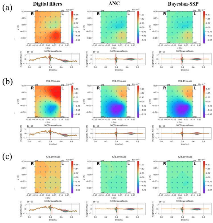
Magnetocardiography (MCG) provides a non-invasive, contactless technique for evaluating the magnetic fields generated by cardiac electrical activity, offering unique spatial insights into cardiac electrophysiology. However, conventional MCG systems depend on superconducting quantum interference devices that require cryogenic cooling and magnetic shielded environments, posing considerable impediments to widespread clinical adoption. In this study, we present a novel MCG system utilizing a high-sensitivity, wide-dynamic-range magnetoresistive sensor array operating at room temperature. To mitigate environmental interference, identical sensors were deployed as reference channels, enabling adaptive noise cancellation (ANC) without the need for traditional magnetic shielding. MCG recordings were obtained from 40 healthy participants, with signals processed using ANC, R-peak-synchronized averaging, and Bayesian spatial signal separation. This approach enabled the reliable detection of key cardiac components, including P, QRS, and T waves, from the unshielded MCG recordings. Our findings underscore the feasibility of a cost-effective, portable MCG system suitable for clinical settings, presenting new opportunities for noninvasive cardiac diagnostics and monitoring.

Keywords: magnetocardiography; medical sensing; noise reduction; signal processing.


Conflict of interest statement

Yu Natsume and Tomohiko Shibuya were employed by the company TDK Corporation. The remaining authors declare that the research was conducted in the absence of any commercial or financial relationships that could be construed as a potential conflict of interest.

Figures

Figure 1
Figure 1
Magnetoresistive sensors and the MCG system. (a) Illustration of the STORM system; (b) configuration of the sensor array; and (c) appearance during measurement.
Figure 2
Figure 2
A diagram illustrating procedures for signal processing.
Figure 3
Figure 3
Noise reduction effect by adaptive noise cancellation (ANC). (a) Noise spectral density before (blue line) and after (red line) application of ANC. The application of ANC reduced the environmental noise level throughout the whole frequency range. (b) The waveform measures environmental signals from one representative channel. The waveform before applying ANC shows alternation in signals (left). The application of ANC eliminates most signals, resulting in the flat waveform (right).
Figure 4
Figure 4
Effect of noise reduction in averaged MCG waveforms after ANC. Waveform of MCG in representative 1 channel (ch.9 of ST-3) is shown after digital filters only (a), after application of ANC and signal averaging (b), and further application of Bayesian SSP (c). (df) The overlapping waveforms of all 42 channels are shown in (ac). The waveforms from each channel are drawn in different colors. (g) Waveform of lead II ECG recorded simultaneously with the MCG.
Figure 5
Figure 5
Waveform array of MCG after the noise reduction process. (a) Waveforms of each magnetoresistive sensor are displayed in the configuration of the sensor array. (b,c) Enlarged waveforms of representative channels selected from the waveform array. Waveforms of Ch. 9 (b) and Ch. 28 (c) are exhibited. The component of the wave (P, Q, R, S, and T) is indicated in each figure.
Figure 6
Figure 6
Isomagnetic field maps after noise reduction processes. Isomagnetic field maps are shown at the timing of the Q-waves (a), R-waves (b), and the end of the QRS complex (c). Each image was constructed using MCG waveforms after digital filters (left), application of ANC and signal averaging (center), and further application of Bayesian SSP (right), respectively.

References

    1. Glaser R. Biophysics: An Introduction. 2nd ed. Springer Science & Business Media; Berlin/Heidelberg, Germany: 2012.
    1. Zhu K., Kiourti A. Real-time magnetocardiography with passive miniaturized coil array in earth ambient field. Sensors. 2023;23:5567. doi: 10.3390/s23125567. - DOI - PMC - PubMed
    1. Brisinda D., Fenici P., Fenici R. Clinical magnetocardiography: The unshielded bet-past, present, and future. Front. Cardiovasc. Med. 2023;10:1232882. doi: 10.3389/fcvm.2023.1232882. - DOI - PMC - PubMed
    1. Xiao W., Sun C., Shen L., Feng Y., Liu M., Wu Y., Liu X., Wu T., Peng X., Guo H. A movable unshielded magnetocardiography system. Sci. Adv. 2023;9:eadg1746. doi: 10.1126/sciadv.adg1746. - DOI - PMC - PubMed
    1. Hu Z., Ye K., Bai M., Yang Z., Lin Q. Solving the magnetocardiography forward problem in a realistic three-dimensional heart-torso model. IEEE Access. 2021;9:107095–107103. doi: 10.1109/ACCESS.2021.3098925. - DOI

Grants and funding

LinkOut - more resources