Review
. 2025 Aug 5;25(15):4812.
doi: 10.3390/s25154812.

Advances in Photoacoustic Imaging of Breast Cancer

Affiliations
Review

Advances in Photoacoustic Imaging of Breast Cancer

Yang Wu et al. Sensors (Basel). .

Abstract

Breast cancer is the leading cause of cancer-related mortality among women world-wide, and early screening is critical for improving patient survival. Medical imaging plays a central role in breast cancer screening, diagnosis, and treatment monitoring. However, conventional imaging modalities-including mammography, ultrasound, and magnetic resonance imaging-face limitations such as low diagnostic specificity, relatively slow imaging speed, ionizing radiation exposure, and dependence on exogenous contrast agents. Photoacoustic imaging (PAI), a novel hybrid imaging technique that combines optical contrast with ultrasonic spatial resolution, has shown great promise in addressing these challenges. By revealing anatomical, functional, and molecular features of the breast tumor microenvironment, PAI offers high spatial resolution, rapid imaging, and minimal operator dependence. This review outlines the fundamental principles of PAI and systematically examines recent advances in its application to breast cancer screening, diagnosis, and therapeutic evaluation. Furthermore, we discuss the translational potential of PAI as an emerging breast imaging modality, complementing existing clinical techniques.

Keywords: breast cancer; diagnostic accuracy; early screening; photoacoustic imaging; therapeutic evaluation.


Conflict of interest statement

The authors declare no conflicts of interest.

Figures

Figure 1
Figure 1
Bed-based whole-breast PAI systems and representative images. (a) The SBH-PACT system and imaging results [48]: (i) System cutaway view (left) and perspective view (right); (ii) From left to right: mammogram, depth-encoded PA image, sagittal PA image, PA image with vascular density overlay, and photoacoustic elastography image. (b) Bed-based 3D-PACT system [51]: (i) System cross-section view after removing the imaging platform; (ii) 3D PA breast images of a subject [from left to right, vascular projection image of the breast (top) and vascular projection image of the breast lateral view (bottom), and cross-sectional image at different coronal planes from the nipple to the chest wall. Each cross-sectional image represents a projection of a 1 cm-thick slice of the breast.]. (c) PAM-02 system and imaging results [54]: (i) Photograph of the system; (ii) US image from the system; (iii) PA structural image; (iv) PA functional image. (d) PAM-03 system and representative images [55]: (i) Photograph of the system and transducer array layout; (ii) and (iii) Imaging results from two healthy volunteers. Both of them are coronal views and colored the signals according to the depth using the color chart. (e) PAI-04 system and PA images [56]: (i) Photograph of the system; (ii) and (iii) PA images of breasts from two healthy participants. Waite rows mean the measured positions for the reproducibility evaluation of the S-factor in the breast. The measured ranges of the S-factor are indicated by the yellow dotted lines. Through measurements, it is possible to distinguish between arteries and veins in the breast. (f) PAM 2 system and imaging results [57]: (i) Photo of the system (left) and imaging tank (right); (ii) Color local maximum intensity projections (LMIPs) of the left breasts from two healthy volunteers acquired at 755 nm, showing sagittal (left, max depth 110 mm) and transverse (right, max depth 100 mm) views. Green crosses follow a blood vessel at a depth of 22 mm from the skin surface. (g) PAM3 breast imaging system and imaging results [58]: (i) Schematic of the PA-US imaging system and the top-view of imaging bowl. (ii) Cross-sectional view of the system core. (iii) Full-SOS-compensated maximum intensity projection (MIP) images in the anterior–posterior, medial–lateral, and cranial–caudal views of the right breast of a healthy volunteer.
Figure 2
Figure 2
Handheld PAI systems based on localized detection and corresponding breast imaging results. (a) Imagio® system and imaging results [64]: (i) Photograph of the Imagio® system; (ii) and (iii) respectively show the US image (left) and the fused PA-US image (right) of a patient with triple-negative invasive ductal carcinoma. (b) The MSOT system schematic and imaging results [65]: (i) System diagram and probe photo; (ii) Schematic of breast tissue structure; (iii) MOST imaging of a healthy breast, revealing the layered structure of the breast. (c) Handheld device developed from a clinical ultrasound platform and imaging results [66]: (i) US/PA dual-modality system schematic; (ii) Handheld probe illustration; (iii) PA imaging results from a patient with a breast tumor; (d) The 3D multispectral PAI system and imaging results [67]: (i) Imaging and probe schematic (enlarged view); (ii) Maximum intensity projections of the breast from a healthy volunteer along the z and x directions with depth-coded color. (e) The Acuity Echo® and PA images [68]: (i) System diagram during acquisition; (ii) PA images of three breast cancer patients, with white contours indicating tumor regions. (f) Schematic of handheld US-PA probe and functional results [69]: (i) System diagram; (ii) PA image (left) showing total hemoglobin (HbT) distribution and fused image (right) displaying lipid (green) and collagen (magenta); blue represents veins with low sO2, red indicates arteries with normal saturation.
Figure 3
Figure 3
PAI performance in noninvasive breast cancer diagnosis across multiple studies. (a) (i) and (ii) Photoacoustic (PA) images acquired using the PAM 2 system, showing transverse and sagittal views of a patient diagnosed with mucinous carcinoma [61] (the dotted line area is the tumor location). (b) Typical diagnostic cases assisted by the ResAM50 deep learning model [86]: (i) A 34-year-old female with a breast lesion detected on US. PA-US imaging showed oxygenation signals around the lesion but none inside. Pathology confirmed a fibroadenoma; (ii) A 64-year-old female with a lesion seen on US. PA-US imaging revealed oxygenation signals both inside and around the lesion. Pathology confirmed invasive ductal carcinoma. (c) Learning-based classification, localization, and segmentation of breast lesions [90]. (i) Each whole-breast PACT image of the patient was rendered as a 2D MAP and divided into four quadrants. (ii) Image processing, feature extraction, and lesion classification and segmentation. (d) Imaging results of a breast cancer patient [56]: (i) A fused image of PA and 3D ultrasound (3D-US). The US data were colored red. Angiogenesis were occurred near the tumor; (ii) A fusion of the S-factor image and 3D-US image (red). The nipple appears light blue due to the spectral similarity between melanin absorption and low sO2. The S-factor indicates the correlation between true and measured sO2. (e) PA-US fusion images [64]: Imagio® system image showing high deoxyhemoglobin levels inside the tumor. Rich vasculature is also visible at the tumor margin (arrow), including radial arteries (green) and veins (red); (f) An image from a handheld PA-US system, showing breast tumor sO2 mapping [66]. (g) Imaging of an inflammatory breast cancer case using the PAM-03 prototype demonstrated a lesion with a diameter of 47 mm [55]: (i) Contrast-enhanced MRI with the lesion circled in red; (ii) Original PA image; (iii) Fused image of the PA image (cyan) and the MRI image (red).
Figure 4
Figure 4
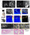
Evaluation of PAI therapeutic efficacy in breast tumors. (a) Comparison of the images acquired by the SBH-PACT and contrast-enhanced MRI [110]. (i) and (ii) represent the PACT images (left, imaging time 15 s without contrast injection) and the contrast-enhanced MRI images (right) of the same breast before (T1) and after (T3) neoadjuvant chemotherapy, respectively. Related structures are marked with white arrows. (b) Dual-modal PA-US imaging system and sentinel lymph node (SLN) imaging results [113]: (i) Photograph of the dual-modal PA-US imaging system; (ii) Photograph of the handheld probe; (iii) Real-time in vivo imaging during SLN biopsy: US image with the lymph node and needle, showing low contrast of them (left); The PA image with the SLN and needle, which are clearly visualized (middle); The co-registered PA-US image with the alignment of the SLN and needle (right); (iv) In vivo imaging of methylene blue and its differentiation from blood vessels: PA image at 650 nm with signals from both methylene blue and hemoglobin (left); PA image at 1064 nm with hemoglobin signals (middle); Image highlighting the distribution of methylene blue (right). (c) Images of a patient breast tumor [44]. Top tow: The UV-PAM images of a unsliced breast tumor and magnified views of the boxed areas. Bottom row: H&E-stained histologic image of the same regions in the breast tumor and magnified views of the boxed areas. The blue dotted line represents the boundary between normal tissue and tumor.

Similar articles

  • Prescription of Controlled Substances: Benefits and Risks.
    Preuss CV, Kalava A, King KC. Preuss CV, et al. 2025 Jul 6. In: StatPearls [Internet]. Treasure Island (FL): StatPearls Publishing; 2025 Jan–. 2025 Jul 6. In: StatPearls [Internet]. Treasure Island (FL): StatPearls Publishing; 2025 Jan–. PMID: 30726003 Free Books & Documents.
  • Management of urinary stones by experts in stone disease (ESD 2025).
    Papatsoris A, Geavlete B, Radavoi GD, Alameedee M, Almusafer M, Ather MH, Budia A, Cumpanas AA, Kiremi MC, Dellis A, Elhowairis M, Galán-Llopis JA, Geavlete P, Guimerà Garcia J, Isern B, Jinga V, Lopez JM, Mainez JA, Mitsogiannis I, Mora Christian J, Moussa M, Multescu R, Oguz Acar Y, Petkova K, Piñero A, Popov E, Ramos Cebrian M, Rascu S, Siener R, Sountoulides P, Stamatelou K, Syed J, Trinchieri A. Papatsoris A, et al. Arch Ital Urol Androl. 2025 Jun 30;97(2):14085. doi: 10.4081/aiua.2025.14085. Epub 2025 Jun 30. Arch Ital Urol Androl. 2025. PMID: 40583613 Review.
  • Photoacoustic imaging for cutaneous melanoma assessment: a comprehensive review.
    Fakhoury JW, Lara JB, Manwar R, Zafar M, Xu Q, Engel R, Tsoukas MM, Daveluy S, Mehregan D, Avanaki K. Fakhoury JW, et al. J Biomed Opt. 2024 Jan;29(Suppl 1):S11518. doi: 10.1117/1.JBO.29.S1.S11518. Epub 2024 Jan 12. J Biomed Opt. 2024. PMID: 38223680 Free PMC article.
  • Pediatric Diffuse High-Grade Gliomas: A Comprehensive Review Of Ad-vanced Methods Of Diagnosis And Treatment.
    Budha RR, Khan SW, Sharma DC, Kulchandani Y, Gsn KR. Budha RR, et al. Curr Cancer Drug Targets. 2025 Jun 30. doi: 10.2174/0115680096365252250618115641. Online ahead of print. Curr Cancer Drug Targets. 2025. PMID: 40598730
  • Short-Term Memory Impairment.
    Cascella M, Al Khalili Y. Cascella M, et al. 2024 Jun 8. In: StatPearls [Internet]. Treasure Island (FL): StatPearls Publishing; 2025 Jan–. 2024 Jun 8. In: StatPearls [Internet]. Treasure Island (FL): StatPearls Publishing; 2025 Jan–. PMID: 31424720 Free Books & Documents.

References

    1. Ren W., Chen M., Qiao Y., Zhao F. Global Guidelines for Breast Cancer Screening: A Systematic Review. Breast. 2022;64:85–99. doi: 10.1016/j.breast.2022.04.003. - DOI - PMC - PubMed
    1. Kim J., Harper A., McCormack V., Sung H., Houssami N., Morgan E., Mutebi M., Garvey G., Soerjomataram I., Fidler-Benaoudia M.M. Global Patterns and Trends in Breast Cancer Incidence and Mortality across 185 Countries. Nat. Med. 2025;31:1154–1162. doi: 10.1038/s41591-025-03502-3. - DOI - PubMed
    1. Beckmann M., Niederacher D., Schnürch H.-G., Gusterson B.A., Bender H.G. Multistep Carcinogenesis of Breast Cancer and Tumour Heterogeneity. J. Mol. Med. 1997;75:429–439. doi: 10.1007/s001090050128. - DOI - PubMed
    1. Nik-Zainal S., Davies H., Staaf J., Ramakrishna M., Glodzik D., Zou X., Martincorena I., Alexandrov L.B., Martin S., Wedge D.C. Landscape of Somatic Mutations in 560 Breast Cancer Whole-Genome Sequences. Nature. 2016;534:47–54. doi: 10.1038/nature17676. - DOI - PMC - PubMed
    1. Nolan E., Lindeman G.J., Visvader J.E. Deciphering Breast Cancer: From Biology to the Clinic. Cell. 2023;186:1708–1728. doi: 10.1016/j.cell.2023.01.040. - DOI - PubMed

LinkOut - more resources