. 2024 Dec;4(4):49.
doi: 10.20517/mtod.2024.59. Epub 2024 Dec 10.

Insulin increases type I collagen synthesis in hepatic stellate cells via α5β1 integrin

Affiliations

Insulin increases type I collagen synthesis in hepatic stellate cells via α5β1 integrin

Milan Dodig et al. Metab Target Organ Damage. 2024 Dec.

Abstract

Aim: A direct effect of insulin on the synthesis of extracellular matrix proteins has been described in extrahepatic organs. The current study investigates the role of insulin in type I collagen production in hepatic stellate cells (HSCs).

Methods: Primary HSC cultures from wild-type mice and from L-SACC1 transgenic mice that exhibit hyperinsulinemia and resultant insulin resistance due to a defect in hepatic insulin clearance were used.

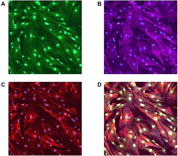
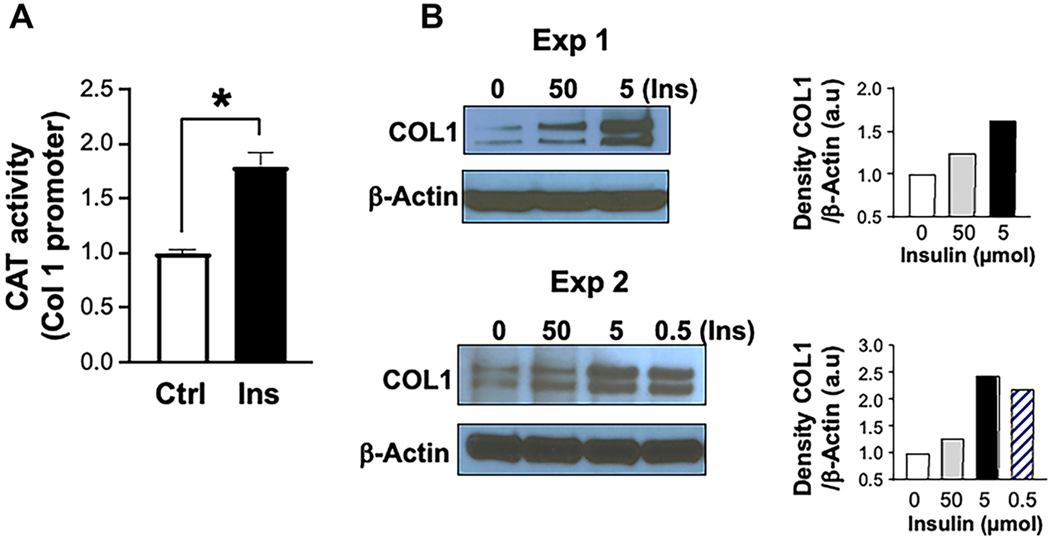
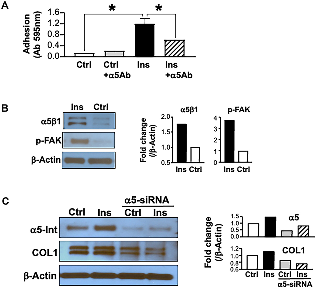
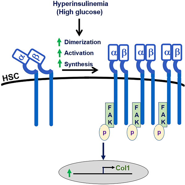
Results: Insulin significantly increased type I collagen synthesis in HSC primary cultures in the presence of high but not low glucose concentrations. Although HSCs contain a functional, insulin-activated PI3 kinase signaling pathway, insulin increases type I collagen synthesis by mechanisms independent of PI3 kinase. Insulin stimulated α5β1 integrin levels and phosphorylation of focal adhesion kinase, a major signaling mediator in the integrin pathway. In addition, α5β1 integrin siRNA interference abolished insulin-mediated type I collagen synthesis by HSCs. L-SACC1 mice showed increased hepatic collagen deposition as compared to wild-type mice. HSCs isolated from L-SACC1 mice synthesize more type I collagen and α5β1 integrin than HSCs isolated from wild-type controls.

Conclusion: Insulin exerts a direct profibrotic impact on HSCs by an α5β1 integrin-mediated mechanism, independently of the PI3 kinase signaling pathway. Thus, chronic hyperinsulinemia may potentiate liver collagen deposition in insulin resistance states. This likely increases the risk of significant fibrosis burden in chronic liver disease associated with insulin resistance.

Keywords: Collagen; hepatic stellate cells; insulin; insulin resistance; α5β1 integrin.


Conflict of interest statement

Conflicts of interest Najjar SM and Kasumov T are Editorial Board members of the journal Metabolism and Target Organ Damage. Najjar SM and Kasumov T were not involved in any steps of editorial processing, notably including reviewers’ selection, manuscript handling and decision making. The other authors declare that there are no conflicts of interest.

Figures

Figure 1.
Figure 1.. Purity of primary hepatic stellate cell cultures.
Primary HSC cultures on day 7 were immunostained for markers of stellate cell phenotype: GFAP (A), α-SMA (B) and nestin (C). The composite image (D) of the triple-stained slide shows that HSCs were stained by all three markers, indicating their purity. HSCs: Hepatic stellate cells; GFAP: glial fibrillary acidic protein; α-SMA: alpha-smooth muscle actin.
Figure 2.
Figure 2.. The insulin effect on type 1 collagen production.
(A) represents higher type I collagen (COL1) in the presence of insulin (Ins, 5 μmol) relative to its absence (Ctrl). Results are compiled from 4 independent experiments. Average Ctrl CAT activity is shown as 1 (100%), with bars representing SEM; (B) represents Western blot analysis of COL1 protein levels following treatment with Ins from two independent experiments. HSC cultures were treated with different doses of Ins ranging from 0.5–50 μmol. All tested doses increased the amount of synthesized COL1, with 5 μmol being the most effective. β-actin was used for normalization against loaded proteins. Gels were scanned using image J v1.53t, and the density of each test band was divided by its corresponding loading Ctrl, represented in arbitrary units (a.u) in the accompanying graph. CAT: Chloramphenicol acetyltransferase; HSCs: hepatic stellate cells; COL1: type I collagen; Ins: insulin; Ctrl: control; SEM: standard error of mean.
Figure 3.
Figure 3.. The effect of glucose and insulin on type I collagen synthesis and HSCs proliferation.
Primary HSC cultures were incubated in DMEM enriched with 50 or 450 mg/dL glucose with (Ins, 5 μmol) or without (Ctrl) Ins. COL1 promoter activity was assayed as in the legend in Figure 2A (A). Promoter activity of COL1 was assayed in cells incubated with different glucose concentrations (in mg/dL, X-axis) without concomitant Ins treatment (B). Alamar blue assay was used to test the effect of Ins on cell proliferation. Cell proliferation was determined by a colorimetric change in the medium measured as a difference in culture media absorbance at 570 and 600 nm (C). HSCs: Hepatic stellate cells; DMEM: Dulbecco’s modified Eagle Medium; COL1: type I collagen; Ins: insulin; Ctrl: control.
Figure 4.
Figure 4.. Insulin signaling in primary HSCs.
(A) depicts AKT phosphorylation in response to insulin. Cells were treated with serum-free medium from days 5–7. On the 7th day, HSCs were treated with Ins (5 μmol) and the proteins were extracted at 15, 30, and 60 min of treatment. Western blot analysis of phosphorylated AKT (normalized to total AKT) showed induced AKT phosphorylation by Ins in HSCs. β-actin was used to demonstrate equal protein loading. Gels were scanned and the density of each band was divided by its corresponding loading Ctrl, and represented as fold change relative to time 0 in the accompanying graph; (B) demonstrates the presence of IRS1 in activated primary HSC cultures, detected by double immunofloresence. The last image shows the co-staining of IRS1 with α-SMA. HSCs: Hepatic stellate cells; AKT: protein kinase B; IRS1: insulin receptor substrate 1; α-SMA: alpha-smooth muscle actin; Ins: insulin; Ctrl: control.
Figure 5.
Figure 5.. Insulin-induced collagen synthesis by HSCs is not mediated by the PI3 Kinase signaling pathway.
HSCs were isolated from transgenic mice livers and plated as described previously. Cells were grown in high-glucose medium with or without Ins (5 μmol) and PI3 kinase inhibitor wortmannin (W) at a dose of 10 μmol/l. Cells were then harvested for protein extraction and CAT assay on day 7. (A) shows Western blot analysis of AKT phosphorylation by Ins in the absence but not in the presence of wortmannin. Gels were scanned and the density of the test band was divided by its corresponding loading Ctrl, represented as a fold change relative to Ctrl in the accompanying graph; (B) represents the CAT assay on day 7, showing no effect of wortmannin on Ins stimulation of COL1 promoter activity. HSCs: Hepatic stellate cells; CAT: chloramphenicol acetyltransferase; AKT: protein kinase B; Ctrl: control; Ins: insulin; COL1: type I collagen.
Figure 6.
Figure 6.. Insulin stimulates α5β1 integrin and its signaling pathway.
(A) represents the adhesion assay in primary cultures of HSCs. Ins treatment (5 μmol) significantly increased HSCs adhesion to fibronectin and pretreating with α5Ab countered this effect, suggesting that increased adhesion is mediated by activation of α5β1 integrin; (B) shows Western blot analysis demonstrating increased levels of α5β1 integrin and p-FAK in Ins-treated HSCs; (C) shows that α5 siRNA inhibits the Ins effect on collagen synthesis in HSCs. Following siRNA-mediated downregulation of α5 integrin, cultures were grown in serum-free high glucose medium with Ins. Western blot analysis shows that Ins stimulation of both α5 integrin and COL1 synthesis was blocked in the absence of α5 integrin. HSCs: Hepatic stellate cells; Ins: insulin; α5Ab: α5β1 integrin blocking antibody; p-FAK: phosphorylated form of focal adhesion kinase; COL1: type I collagen.
Figure 7.
Figure 7.. Type 1 collagen expression is increased in the livers of L-SACC1 transgenic mice.
Sirius red staining (visualized under polarized light) of representative liver sections from wild-type (A and B) and L-SACC1 mice (C and D). L-SACC1 livers contain more stained birefringent COL1 fibers (seen as yellow on the black background). COL1: type I collagen.
Figure 8.
Figure 8.. Elevated Type I collagen and α5β1 integrin levels in primary HSCs isolated from L-SACC1 mice.
HSCs were isolated as described in Methods and cultured in DMEM with 450 mg/dL glucose, 10% FCS, and 10% HS. Cells were harvested at times indicated on individual panels for Western blot analysis and analyzed by Alamar blue assay for proliferation on day 6. (A) shows Western blot analysis demonstrating increased production of COL1 and α5 integrin by HSCs isolated from L-SACC1 mice compared to cells from wild-type animals; (B) represents a Western blot analysis on days 3, 5, and 7, showing significantly higher COL1 levels in L-SACC1 than in wild-type mice only on day 7. This suggests that HSCs from L-SACC1 mice do not undergo activation earlier in culture. In contrast, α5 integrin levels are increased even after 3 days in culture, with a more pronounced increase in HSCs from L-SACC1 compared to those from wild-type mice; (C) shows no difference in HSC proliferation in primary culture between L-SACC1 and wild-type cells. Cell proliferation was determined by a colorimetric change in the medium measured as a difference in culture media absorbance at 570 and 600 nm. HSCs: Hepatic stellate cells; DMEM: Dulbecco’s modified Eagle Medium; COL1: type I collagen.
Figure 9.
Figure 9.. Proposed model of the effect of hyperinsulinemia and hyperglycemia on α5β1 integrin signaling and type I collagen synthesis.
HSCs: Hepatic stellate cells; FAK: focal adhesion kinase.

Similar articles

References

    1. Gastaldelli A, Cusi K. From NASH to diabetes and from diabetes to NASH: mechanisms and treatment options. JHEP Rep 2019;1:312–28. - PMC - PubMed
    1. El-Serag HB, Tran T, Everhart JE. Diabetes increases the risk of chronic liver disease and hepatocellular carcinoma. Gastroenterology 2004;126:460–8. - PubMed
    1. Fujii H, Kawada N; Japan Study Group Of Nafld Jsg-Nafld. The role of insulin resistance and diabetes in nonalcoholic fatty liver disease. Int J Mol Sci 2020;21:3863. - PMC - PubMed
    1. Bugianesi E, Marchesini G, Gentilcore E, et al. Fibrosis in genotype 3 chronic hepatitis C and nonalcoholic fatty liver disease: role of insulin resistance and hepatic steatosis. Hepatology 2006;44:1648–55. - PubMed
    1. Tomic D, Shaw JE, Magliano DJ. The burden and risks of emerging complications of diabetes mellitus. Nat Rev Endocrinol 2022;18:525–39. - PMC - PubMed

LinkOut - more resources