

Evaluation of the Effect of Tranexamic Acid on Meniscus Healing and Articular Cartilage in a Rabbit Model
- PMID: 40808776
- PMCID: PMC12339491
- DOI: 10.1177/15563316251357603
Evaluation of the Effect of Tranexamic Acid on Meniscus Healing and Articular Cartilage in a Rabbit Model
Abstract
Background: The formation of a stable fibrin clot plays an important role in early tissue repair. Tranexamic acid (TXA), a potent fibrinolysis inhibitor, prevents fibrin clot dissolution.
Purpose: We sought to test the effect of intra-articular TXA administration on meniscus healing and articular cartilage status in a rabbit model.
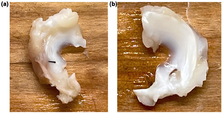
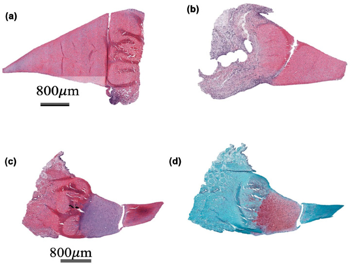
Methods: Thirty-two rabbits underwent bilateral knee surgery with creation of a 1.5-mm circular defect in the anterior horn of the lateral meniscus and a 3-mm longitudinal tear with repair in the anterior horn of the medial meniscus. Twelve rabbits were used for an initial TXA dose determination study. Twenty rabbits were then injected with 50 mg/mL of TXA in the left knee while the right knee served as a control. Animals were sacrificed at 2-, 4-, and 8-week timepoints. Eight rabbits underwent biomechanical analysis. Semiquantitative histological analysis compared meniscal healing and articular cartilage between TXA-treated and control knees.
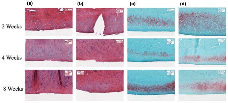
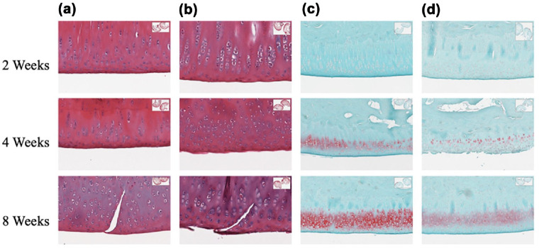
Results: Both circular defects of the lateral meniscus and longitudinal tear injuries of the medial meniscus showed no difference in healing across all timepoints. At 2 weeks post-surgery, TXA-treated knees exhibited reduced tibial articular cartilage structure compared to controls. By week 8, control knees had higher proteoglycan content in all femoral articular cartilage zones compared to TXA-treated knees. Biomechanical analysis was inconclusive.
Conclusion: This rabbit study found that TXA administration did not enhance healing following meniscus repair. Moreover, intra-articular TXA appeared to have exerted an adverse effect on articular cartilage, possibly due to the detrimental effects of persistent blood in a joint. Further studies will be critically important to determine the effect of TXA administration at various time points after surgical repair.
Keywords: articular cartilage; fibrin clot; meniscus; rabbit; tranexamic acid (TXA).
© The Author(s) 2025.
Conflict of interest statement
The author(s) declared the following potential conflicts of interest with respect to the research, authorship, and/or publication of this article: Scott Rodeo, MD, reports relationships with Arthritis Foundation, OREF, Ortho RTI, NIH, Angiocrine Biosciences, Novartis, Advance Medical, Jannu Therapeutics, and Overture Medical. The other authors declare no potential conflicts of interest.
Figures
Similar articles
-
Pharmacological interventions for the prevention of bleeding in people undergoing elective hip or knee surgery: a systematic review and network meta-analysis.Cochrane Database Syst Rev. 2024 Jan 16;1(1):CD013295. doi: 10.1002/14651858.CD013295.pub2. Cochrane Database Syst Rev. 2024. PMID: 38226724 Free PMC article.
-
Population Pharmacokinetics of Intra-articular and Intravenous Administration of Tranexamic Acid in Patients Undergoing Total Knee Replacement.Clin Pharmacokinet. 2022 Jan;61(1):83-95. doi: 10.1007/s40262-021-01043-9. Epub 2021 Jul 13. Clin Pharmacokinet. 2022. PMID: 34255299
-
Can the Acetabular Labrum Be Reconstructed With a Meniscal Allograft? An In Vivo Pig Model.Clin Orthop Relat Res. 2024 Feb 1;482(2):386-398. doi: 10.1097/CORR.0000000000002860. Epub 2023 Sep 19. Clin Orthop Relat Res. 2024. PMID: 37732715 Free PMC article.
-
Intra-articular corticosteroid for knee osteoarthritis.Cochrane Database Syst Rev. 2015 Oct 22;2015(10):CD005328. doi: 10.1002/14651858.CD005328.pub3. Cochrane Database Syst Rev. 2015. PMID: 26490760 Free PMC article.
-
Sertindole for schizophrenia.Cochrane Database Syst Rev. 2005 Jul 20;2005(3):CD001715. doi: 10.1002/14651858.CD001715.pub2. Cochrane Database Syst Rev. 2005. PMID: 16034864 Free PMC article.
References
-
- Alvarez JC, Santiveri FX, Ramos I, Vela E, Puig L, Escolano F. Tranexamic acid reduces blood transfusion in total knee arthroplasty even when a blood conservation program is applied. Transfusion. 2008;48(3):519–525. - PubMed
-
- Ambra LF, de Girolamo L, Niu W, Phan A, Spector M, Gomoll AH. No effect of topical application of tranexamic acid on articular cartilage. Knee Surg Sports Traumatol Arthrosc. 2019;27(3):931–995. - PubMed
-
- Arnoczky SP, Warren RF, Spivak JM. Meniscal repair using an exogenous fibrin clot. An experimental study in dogs. J Bone Joint Surg Am. 1988;70(8):1209–1217. - PubMed
-
- Blanco FJ, Guitian R, Vázquez-Martul E, de Toro FJ, Galdo F. Osteoarthritis chondrocytes die by apoptosis: a possible pathway for osteoarthritis pathology. Arthritis Rheum. 1998;41(2):284–289. - PubMed
-
- Bolam SM, O’Regan-Brown A, Paul Monk A, Musson DS, Cornish J, Munro JT. Toxicity of tranexamic acid (TXA) to intra-articular tissue in orthopaedic surgery: a scoping review. Knee Surg Sports Traumatol Arthrosc. 2021;29(6):1862–1871. - PubMed
LinkOut - more resources
Full Text Sources
