

Challenges and opportunities for new intraoperative optical techniques in the surgical treatment of pituitary adenomas: a review
- PMID: 40808829
- PMCID: PMC12344518
- DOI: 10.1117/1.JBO.30.8.080901
Challenges and opportunities for new intraoperative optical techniques in the surgical treatment of pituitary adenomas: a review
Abstract
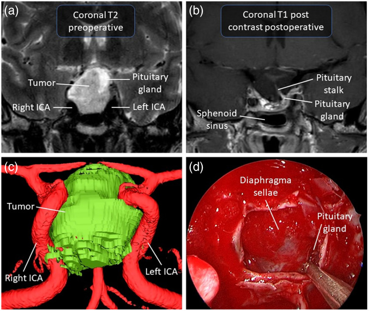
Significance: Surgery is a common intervention for patients with pituitary adenomas, particularly those experiencing endocrine symptoms or mass effect. Persistent challenges in pituitary surgery include the detection of small microadenomas, difficulty in discerning residual tumor from normal gland, and infiltrative adenomas. Although standard perioperative diagnostics include magnetic resonance imaging (MRI), computed tomography, ultrasound imaging, and neuronavigation, some centers employ intraoperative MRI, ultrasound, and fluorescence-guided endoscopy to increase the rate of gross total resection and preserve pituitary function. However, these techniques are often limited by availability, time requirements, cost, and inability to provide histological diagnosis.
Aim: This review addresses opportunities to optimize both the extent of resection and gland preservation in pituitary adenoma procedures. We discuss the existing constraints faced in pituitary surgery and showcase the current and emerging detection techniques employed in clinical practice, as well as their limitations. We also discuss newer probing approaches such as elastography and Raman spectroscopy.
Approach: We outline key attributes for an ideal optical tool, considering surgical theater functionality, ergonomics, and result reliability and accuracy.
Results: A case study is presented describing the recent development of a fiber-optics instrument specifically designed for endonasal applications based on clinical requirements, along with preliminary data supporting the feasibility of intraoperative implementation.
Conclusions: Current imaging and navigation tools, although invaluable, have inherent limitations in resolution, integration, and molecular specificity. Raman spectroscopy offers a promising, label-free method for real-time tissue identification, especially when integrated into fiber-optic probes for endonasal use. As a complementary tool, it could enhance intraoperative decision-making and surgical precision. Further clinical validation is needed to support its integration into standard workflows.
Keywords: Raman spectroscopy; clinical translation; instrumentation; neurosurgery; pituitary adenoma; tissue optics.
© 2025 The Authors.
Figures


References
Publication types
MeSH terms
LinkOut - more resources
Full Text Sources
Medical
Research Materials
