Review
. 2025 Jul 30:18:1642519.
doi: 10.3389/fnmol.2025.1642519. eCollection 2025.

Proteomic insights into the biology of dopaminergic neurons

Affiliations
Review

Proteomic insights into the biology of dopaminergic neurons

Claudia Cavarischia-Rega et al. Front Mol Neurosci. .

Abstract

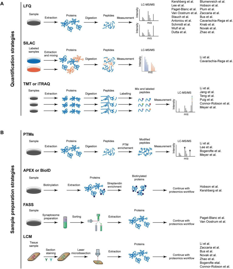
Dopaminergic neurons, primarily located in the substantia nigra, hypothalamus, and ventral tegmental area of the brain, play crucial roles in motor control, reward, motivation, and cognition. Alterations in their function are associated with numerous neurological and psychiatric disorders, such as Parkinson's disease, but also Schizophrenia, substance use disorders, and bipolar disorder. Recent advances in mass spectrometry-based proteomics have enabled the comprehensive profiling of protein expression, turnover, subcellular localization, and post-translational modifications at an unprecedented depth of analysis. This review summarizes the developments in proteomic approaches taken to study dopaminergic neurons. We cover findings from global and spatial proteomics studies that revealed brain region-specific protein signatures, as well as dynamic turnover of proteins and the importance of mitochondrial and synaptic proteins for the health and vulnerability of dopaminergic neurons. Combined with advanced molecular cell biology tools, such as growth in microfluidic devices, fluorescent-activated synaptosome sorting, and enzymatic proximity labeling, modern proteomics allows for investigation of synaptic and subcellular proteomes. Despite these advancements, the complexity of the human brain and its cell-specific characteristics remain a challenge. The continuing integration of advanced proteomic techniques with other -omics will eventually yield improved and mechanistic understanding of dopaminergic neurons in health and disease.

Keywords: dopaminergic neurons; iPSCs; protein turnover; proteomics; synaptosomes.


Conflict of interest statement

The authors declare that the research was conducted in the absence of any commercial or financial relationships that could be construed as a potential conflict of interest.

Figures

Diagram illustrating dopaminergic pathways in the brain. Part A shows pathways including nigrostriatal (red), mesocortical (blue), mesolimbic (purple), and tubero-infundibular (green), labeled with brain regions like striatum and thalamus. Part B depicts a neuron with labeled parts like axon and synapse, highlighting neurotransmission processes, mentioning studies by various authors.
FIGURE 1
Dopamine pathways and subcellular compartments of DA neurons. (A) Schematic of the different brain regions where dopaminergic neurons are located. The four main dopamine pathways (nigrostriatal, mesocortical, mesolimbic, and tuberoinfundibular) are also depicted in different colors (red, purple, blue, and green respectively). (B) DA neuron with a zoom-in of one synapse. The key steps in the synthesis of dopamine and its release can be seen. Papers that are discussed across the review related to axonal proteomics or synaptosomal proteomics are also annotated in the figure.
Diagram showing quantification and sample preparation strategies for proteomics. Section A includes LFQ, SILAC, and TMT/iTRAQ, each describing steps from sample extraction to LC-MS/MS measurement. Section B covers PTMs, APEX/BioID, FASS, and LCM, detailing steps like enrichment and extraction. Both sections list reference studies.
FIGURE 2
Proteomic workflows. (A) The workflows for quantification strategies in proteomics. The upper panel depicts a standard workflow for LFQ, wherein proteins are extracted from a sample and subsequently digested into peptides. Thereafter, the peptides are measured using LC-MS/MS. Each sample is measured independently. The middle panel shows SILAC strategy. In this approach, samples are cultivated in SILAC media, allowing the proteins to incorporate the isotopic labels during their synthesis. Following this, the proteins are extracted, mixed in a 1:1 ratio, and digested into peptides for measurement. The lower panel outlines the workflow for TMT or Isobaric Tags for Relative and Absolute Quantitation (iTRAQ) labeling. In this process, proteins are digested, and the peptides are subsequently labeled. After labeling, the peptides are mixed and measured. Quantitative information is derived from reporter ions in the MS/MS spectrum. (B) The strategies for sample preparation. The upper panel depicts the preparation of samples for post-translational modification (PTM) studies. Proteins are extracted and digested into peptides. Following this, a PTM enrichment step is conducted, during which unmodified peptides are removed. The samples are subsequently measured. In the upper-middle panel, the apex or BioID workflow is presented. The protein of interest, tagged with an enzyme, is activated by the addition of a reagent, resulting in the enzyme biotinylating proteins in its immediate vicinity. Cells are lysed, and the biotinylated proteins are enriched using streptavidin beads. Thereafter, proteins are digested into peptides and further analyzed with LC-MS/MS. The FASS workflow is depicted in the lower-middle panel. Initially, synaptosomes are enriched and their subpopulations are sorted using fluorescent tagging of synaptic markers. Subsequently, proteins are digested and measured. In the lower panel, the LCM workflow is illustrated, where a tissue sample is stained with various antibodies, and a specific region is microdissected and isolated using a laser. This is followed by a standard proteomic workflow. The papers discussed in the review are indicated adjacent to the workflow utilized.

Similar articles

  • Prescription of Controlled Substances: Benefits and Risks.
    Preuss CV, Kalava A, King KC. Preuss CV, et al. 2025 Jul 6. In: StatPearls [Internet]. Treasure Island (FL): StatPearls Publishing; 2025 Jan–. 2025 Jul 6. In: StatPearls [Internet]. Treasure Island (FL): StatPearls Publishing; 2025 Jan–. PMID: 30726003 Free Books & Documents.
  • Short-Term Memory Impairment.
    Cascella M, Al Khalili Y. Cascella M, et al. 2024 Jun 8. In: StatPearls [Internet]. Treasure Island (FL): StatPearls Publishing; 2025 Jan–. 2024 Jun 8. In: StatPearls [Internet]. Treasure Island (FL): StatPearls Publishing; 2025 Jan–. PMID: 31424720 Free Books & Documents.
  • Reduced expression of Pss gene in Drosophila cortex glia causes dopaminergic cell death.
    Pak B, Kim C, Kwon SH, Lee JK, Jeon SH. Pak B, et al. J Parkinsons Dis. 2025 Aug;15(5):957-969. doi: 10.1177/1877718X251349407. Epub 2025 Jun 16. J Parkinsons Dis. 2025. PMID: 40518954
  • The Black Book of Psychotropic Dosing and Monitoring.
    DeBattista C, Schatzberg AF. DeBattista C, et al. Psychopharmacol Bull. 2024 Jul 8;54(3):8-59. Psychopharmacol Bull. 2024. PMID: 38993656 Free PMC article. Review.
  • Management of urinary stones by experts in stone disease (ESD 2025).
    Papatsoris A, Geavlete B, Radavoi GD, Alameedee M, Almusafer M, Ather MH, Budia A, Cumpanas AA, Kiremi MC, Dellis A, Elhowairis M, Galán-Llopis JA, Geavlete P, Guimerà Garcia J, Isern B, Jinga V, Lopez JM, Mainez JA, Mitsogiannis I, Mora Christian J, Moussa M, Multescu R, Oguz Acar Y, Petkova K, Piñero A, Popov E, Ramos Cebrian M, Rascu S, Siener R, Sountoulides P, Stamatelou K, Syed J, Trinchieri A. Papatsoris A, et al. Arch Ital Urol Androl. 2025 Jun 30;97(2):14085. doi: 10.4081/aiua.2025.14085. Epub 2025 Jun 30. Arch Ital Urol Androl. 2025. PMID: 40583613 Review.

References

    1. Aebersold R., Mann M. (2016). Mass-spectrometric exploration of proteome structure and function. Nature 537 347–355. 10.1038/nature19949 - DOI - PubMed
    1. Aguila J., Cheng S., Kee N., Cao M., Wang M., Deng Q., et al. (2021). Spatial RNA sequencing identifies robust markers of vulnerable and resistant human midbrain dopamine neurons and their expression in Parkinson’s disease. Front. Mol. Neurosci. 14:699562. 10.3389/fnmol.2021.699562 - DOI - PMC - PubMed
    1. Alum E. U. (2025). AI-driven biomarker discovery: Enhancing precision in cancer diagnosis and prognosis. Discov. Oncol. 16:313. 10.1007/s12672-025-02064-7 - DOI - PMC - PubMed
    1. Antoniou N., Prodromidou K., Kouroupi G., Boumpoureka I., Samiotaki M., Panayotou G., et al. (2022). High content screening and proteomic analysis identify a kinase inhibitor that rescues pathological phenotypes in a patient-derived model of Parkinson’s disease. NPJ Parkinsons Dis. 8:15. 10.1038/s41531-022-00278-y - DOI - PMC - PubMed
    1. Antonov S., Novosadova E. (2021). Current state-of-the-art and unresolved problems in using human induced pluripotent stem cell-derived dopamine neurons for Parkinson’s disease drug development. Int. J. Mol. Sci. 22:3381. 10.3390/ijms22073381 - DOI - PMC - PubMed

LinkOut - more resources