. 2025 Jul 30:16:1583597.
doi: 10.3389/fimmu.2025.1583597. eCollection 2025.

Unveiling B7/CD28 family proteins in hepatocellular carcinoma: insights into communication and prognostic significance

Affiliations

Unveiling B7/CD28 family proteins in hepatocellular carcinoma: insights into communication and prognostic significance

Yan Cai et al. Front Immunol. .

Abstract

Background: Immunotherapy has made remarkable achievements in cancer treatment, but it still faces the challenge of limited response rates in liver cancer therapy. Although there has been extensive research on the role of programmed cell death-ligand 1 (PD-L1) in hepatocellular carcinoma (HCC), our understanding of the effects of other inhibitory B7/CD28 family members is still limited despite advancements in prognostic tools, more specific, accurate, and robust biomarkers are required to improve HCC patient prognoses.

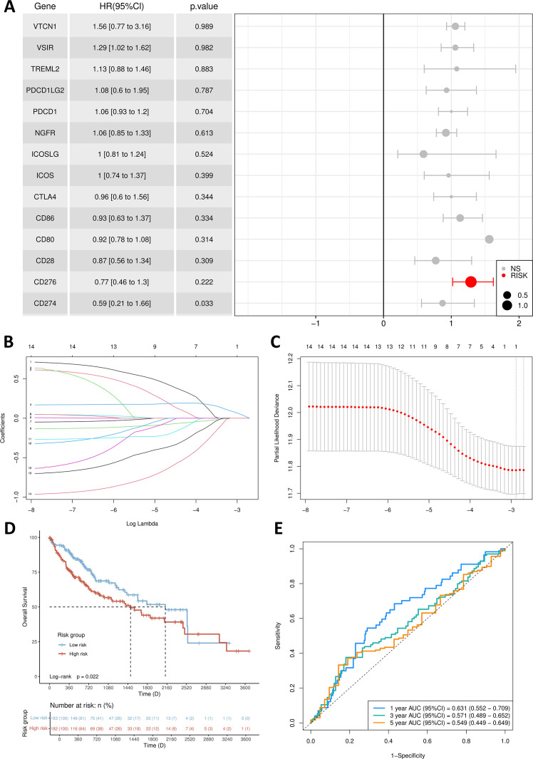
Methods: We acquired the single-cell sequencing data from relevant literature and selected 42 liver tissue samples, including 89,246 cells from HCC patients, to investigate the expression, localization, and intercellular communications of the B7/CD28 family in HCC. Within the Cancer Genome Atlas dataset, we utilized Lasso and Cox regression analyses to develop a risk model for identifying the most pertinent B7/CD28 family proteins associated with prognosis. Subsequently, we conducted a retrospective analysis of 94 HCC patients who underwent hepatectomy at our institution and determined the prognostic significance of this malignancy.

Results: Based on the single-cell RNA sequencing data, we have delineated various members of the B7/CD28 family and their corresponding receptors. We have elucidated their distribution on tumor cells and immune cells, revealing active intercellular communications among tumor cells, fibroblasts, and epithelial cells. Through the implementation of Lasso, we have pinpointed a significant correlation between the B7H3 molecule and prognosis. Leveraging multiplex immunofluorescence, we were able to discern three distinct patterns of B7H3 expression-tumor-associated, stroma-associated, and a hybrid form encompassing both. Notably, the presence of B7H3 in the stroma exhibited the most robust correlation with prognosis. Furthermore, the efficacy of our prognostic signature was validated through clinicopathological analyses conducted at our institution.

Conclusions: In conclusion, the B7/CD28 family plays an active role in the tumor immune microenvironment and cellular communication. B7H3 could serve as an indicator for predicting the outcome of HCC. Additional investigation is required to validate these discoveries in future groups of individuals and assess their viability as therapies guided by biomarkers.

Keywords: B7/CD28 family members; B7H3; hepatocellular carcinoma (HCC); immunotherapy; inhibitory B7 family members; single-cell RNA sequencing; tumor immune environment.


Conflict of interest statement

The authors declare that the research was conducted in the absence of any commercial or financial relationships that could be construed as a potential conflict of interest.

Figures

Figure 1
Figure 1
Healthy liver and tumor samples. (A) t-SNE plot of 42 samples. (B) t-SNE plot comparing healthy liver and liver cancer samples. (C) Heatmap showing the expression of marker genes. Red indicates higher expression levels, while blue indicates lower expression levels. (D) Bar graph showing the relative proportions of cell types in healthy liver and liver cancer. Each cell type is represented by a different color. (E) Bubble plot displaying the expression of marker genes in major cell types. The color of the dots reflects the expression level, and the size of the dots represents the percentage of cells expressing the marker gene in different cell types.
Figure 2
Figure 2
Cell-Cell Ligand-Receptor Interactions Predicted by CellChat in B7/CD28 family Protein Tumor Cells, Endothelial Cells, and Immune Cells. (A) The expression of B7/CD28 family proteins in the clusters. (A, B) t-SNE plot of hepatic-derived cells and stroma cells. (C) Bubble plots of the B7/CD28 family protein in different cell types. (D) Heatmap showing the B7/CD28 family gene expressions across the 11 cell types in healthy liver and healthy Liver and tumor Samples. (E) Bubble plot of ligand-receptor pairs for B7/CD28 family proteins. The color of the dots reflects the communication probability, and the size of the dots represents the computed p-value. Each cell group is displayed using a different color. Blank space indicates a communication probability of zero. The p-values are calculated using a two-sided permutation test. (F) Heatmap showing the strength of interactions between healthy liver and liver tumor cells.
Figure 3
Figure 3
Establishment of prognosis signature. (A) Multivariate Cox regression forest plot. (B) B7/CD28 family proteins were analyzed by LASSO regression. (C) The tuning parameter selection of the LASSO analysis. (D) Kaplan-Meier survival curves for the high-risk group and low-risk group. (E) The AUCs for 1-, 3- and 5-year ROC curves.
Figure 4
Figure 4
The expression pattern of B7H3 in tumor tissues. (A) The representative IHC images of Isotype control antibody and B7H3 antibody; (B) The representative IHC images of diffuse HCC and well-circumscribed HCC; (C) Representative multi-immunofluorescence images of AFP (green) and B7H3 (red). The nucleus is labeled with DAPI (blue). (D) Proportions of B7H3 on tumor cells (TCs), in stroma and in both. (E) The representative IHC images of B7H3 from serial sections. (F) OS and PFS for HCC patients based on the expression of B7H3 on tumor tissue. (G) OS and PFS for HCC patients based on the expression of B7H3 in the stroma and on tumor cells.

Similar articles

References

    1. Siegel RL, Giaquinto AN, Jemal A. Cancer statistics, 2024. CA Cancer J Clin. (2024) 74:12–49. doi: 10.3322/caac.21820, PMID: - DOI - PubMed
    1. Liu Y, Xun Z, Ma K, Liang S, Li X, Zhou S, et al. Identification of a tumour immune barrier in the HCC microenvironment that determines the efficacy of immunotherapy. J Hepatol. (2023) 78:770–82. doi: 10.1016/j.jhep.2023.01.011, PMID: - DOI - PubMed
    1. Sangro B, Sarobe P, Hervás-Stubbs S, Melero I. Advances in immunotherapy for hepatocellular carcinoma. Nat Rev Gastroenterol Hepatol. (2021) 18:525–43. doi: 10.1038/s41575-021-00438-0, PMID: - DOI - PMC - PubMed
    1. Tan S, Xu Y, Wang Z, Wang T, Du X, Song X, et al. Tim-3 hampers tumor surveillance of liver-Resident and conventional NK cells by disrupting PI3K signaling. Cancer Res. (2020) 80:1130–42. doi: 10.1158/0008-5472.CAN-19-2332, PMID: - DOI - PubMed
    1. Burke KP, Chaudhri A, Freeman GJ, Sharpe AH. The B7:CD28 family and friends: Unraveling coinhibitory interactions. Immunity. (2024) 57:223–44. doi: 10.1016/j.immuni.2024.01.013, PMID: - DOI - PMC - PubMed

MeSH terms