. 2025 Jul 30:16:1618404.
doi: 10.3389/fimmu.2025.1618404. eCollection 2025.

IL-7 armed binary CAR T cell strategy to augment potency against solid tumors

Affiliations

IL-7 armed binary CAR T cell strategy to augment potency against solid tumors

Alejandro G Torres Chavez et al. Front Immunol. .

Abstract

Introduction: Clinical studies of T cells engineered with chimeric antigen receptor (CAR) targeting CD19 in B-cell malignancies have demonstrated that relapse due to target antigen (CD19) loss or limited CAR T cell persistence is a common occurrence. The possibility of such events is greater in solid tumors, which typically display more heterogeneous antigen expression patterns and are known to directly suppress effector cell proliferation and persistence. T cell engineering strategies to overcome these barriers are being explored. However, strategies to simultaneously address both antigen heterogeneity and T cell longevity, while localizing anti-tumor effects at disease sites, remain limited.

Methods: In this study we explore a dual antigen targeting strategy by directing independent CARs against the solid tumor targets PSCA and MUC1. To enhance functional persistence in a tumor-localized manner, we expressed the transgenic IL-7 cytokine and receptor (IL-7Rα) in respective CAR products.

Results: This binary strategy, which incorporates dual antigen targeting with transgenic cytokine support, resulted in enhanced potency, T cell expansion, and durable antitumor effects in a pancreatic tumor model compared to single antigen targeting or dual antigen targeting in absence of the transgenic cytokine support.

Discussion: The transgenic IL-7 armed binary CAR T cell approach could improve the efficacy of CAR-based therapies for solid tumors.

Keywords: IL-7; IL-7R; MUC 1; PSCA; T-cell therapy; chimeric antigen receptor; pancreatic cancer; solid tumor.


Conflict of interest statement

AL is a co-founder and equity holder for AlloVir and Marker Therapeutics, and was a consultant to AlloVir. JV is the chief executive officer, a co-founder, and equity holder of Marker Therapeutics. The remaining authors declare that the research was conducted in the absence of any commercial or financial relationships that could be construed as a potential conflict of interest.​

Figures

Figure 1
Figure 1
Limited T cell persistence restricts durable tumor control in a dual target setting. (A) Illustrations of the CARs targeting PSCA (top) and MUC1 (bottom) used to generate CAR T cells. (B) PSCA (left) and MUC1 (right) CAR expression in C.P and C.M T cells measured by flow cytometry 3 days post-transduction. Non-transduced (NT) T cells used as negative controls (t-tests, n=3, ns = no significant difference, *p<.05, **p<.01, ***p<.001, ****p<.0001). (C) Detection of PSCA (top) and MUC1 (bottom) expression by CAPAN1 tumor cells measured using flow cytometry. (D) Cytolytic activity of C.P T cells against 293T (PSCA negative control) and CAPAN1 tumor cells measured using 51Cr-release assay at the effector to target ratio (E:T) of 20:1. NT cells used as controls (t-tests, n=3, ns = no significant difference, *p<.05, **p<.01, ***p<.001, ****p<.0001). (E) Anti-tumor activity of C.M T cells against 293T and CAPAN1 cells measured using 51Cr-release assay at the effector to target ratio (E:T) of 20:1 (t-tests, n=3, ns = no significant difference, *p<.05, **p<.01, ***p<.001, ****p<.0001). (F) CAPAN1 tumor luminescence measured by IVIS imaging in a long-term co-culture assay with CAR T cells in G-Rex 6-well plates. Untreated (tumor-only) condition used as control (one-way Anova on day 27, n=3, ns = no significant difference, *p<.05, **p<.01, ***p<.001, ****p<.0001). (G) PSCA (top) and MUC1 (bottom) expression by CAPAN1 cells from the co-culture experiment in (1F), measured on day 20 by flow cytometry. All T cell treated conditions normalized to the control (t-tests, n=3, ns = no significant difference, *p<.05, **p<.01, ***p<.001, ****p<.0001). (H) CAPAN1 luminescence in C.M, C.P, and C.P+C.M treated conditions in (1F) after re-treatment with respective CAR T cells on day 27 (t-tests on day 35, n=3, ns, no significant difference, *p<.05, **p<.01, ***p<.001, ****p<.0001).
Figure 2
Figure 2
Engineering CAR T cells to secrete and efficiently utilize IL-7 cytokine (A). Illustrations of the IL-7 cytokine construct containing mOrange fluorescent protein for transgene detection by flow cytometry (top) and detection of both the PSCA CAR and IL-7 transgene expression in C.P7 T cells from a representative donor after serial transduction (bottom). (B) Summary data indicating PSCA CAR and IL-7 double-transduced T cells compared to NT cells (t-tests, n=3, ns = no significant difference, *p<.05, **p<.01, ***p<.001, ****p<.0001). (C) Production of IL-7 by C.P7 cells after stimulation with irradiated K562 cells engineered to express PSCA, measuring using ELISA (t-tests, n=3, ns = no significant difference, *p<.05, **p<.01, ***p<.001, ****p<.0001). (D) Diagram illustrating the IL-7Rα construct containing GFP for transgene detection (top) and flow cytometry data for a representative donor demonstrating the expression of both the MUC1 CAR and the IL-7Rα transgenes in C.M7R T cells. (E) Summary data comparing IL-7Rα detection by flow cytometry in NT, C.M, and C.M7R cells (one-way ANOVA, n=3, ns = no significant difference, *p<.05, **p<.01, ***p<.001, ****p<.0001). (F) Quantification of C.M and C.M7R T cells using trypan blue exclusion during culture with irradiated CAPAN1 tumor cells in presence or absence of recombinant IL-7 cytokine (t-tests on day 8, n=3, ns, no significant difference, *p<.05, **p<.01, ***p<.001, ****p<.0001).
Figure 3
Figure 3
IL-7 engineered C.P7 T cells support the expansion of C.M7R overexpressing IL-7Rα. (A) Growth of C.M7R T cells in cell culture medium, and conditioned medium collected from C.P or C.P7 T cells stimulated with irradiated CAPAN1 tumor cells. Cells were quantified by manual counting (t-tests on day 5, n=3, ns = no significant difference, *p<.05, **p<.01, ***p<.001, ****p<.0001). (B) Total cell numbers in irradiated CAPAN1-stimulation co-cultures of the indicated T cell types (t-tests on day 14, n=3, ns = no significant difference, *p<.05, **p<.01, ***p<.001, ****p<.0001). (C) Differentiation of C.P7 and C.M7R T cells based on GFP (surrogate for transgenic IL-7Rα) using flow cytometry to quantify the proportion of these cells in the C.P7+C.M7R condition at the start and the end of the stimulation experiment in (3B) (t-tests on day 14, n=3, ns = no significant difference, *p<.05, **p<.01, ***p<.001, ****p<.0001). (D) Flow cytometric quantification of C.M7R T cells cultured with NT (control), C.P, or C.P7 T cells pre-stimulated with irradiated CAPAN1. Arrows indicate time points at which additional pre-stimulated C.P7 T cells were added. Dashed lines indicate conditions which were monitored for an additional time point without the additional dose of pre-stimulated C.P7 T cells (t-tests at indicated time-points, n=3, ns, no significant difference, *p<.05, **p<.01, ***p<.001, ****p<.0001).
Figure 4
Figure 4
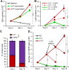
Dual-targeted binary T cells exhibit enhanced anti-tumor activity and persistence. (A) Representative bioluminescent images of GFP/FFLuc expressing CAPAN1-seeded Algimatrix cultures in the long-term co-culture assay using 6-well G-Rex devices. Two replicates shown for each condition (B) Quantification of bioluminescence signal from the CAPAN-1 cells [illustrated in (4A)] during the co-culture experiment under indicated treatment conditions (t-tests on day 20, n=3, ns = no significant difference, *p<.05, **p<.01, ***p<.001, ****p<.0001). (C) Live T cell counts using flow cytometry on day 20 for the different treatment conditions in (B) (one-way ANOVA, n=3, ns, no significant difference, *p<.05, **p<.01, ***p<.001, ****p<.0001).
Figure 5
Figure 5
Binary T cells demonstrate superior anti-tumor activity against pancreatic spheroids. (A) Representative bioluminescent images of GFP/FFLuc expressing CAPAN1 tumor cells generated spheroids during long-term co-culture. Three replicates illustrated for each condition. (B) Quantitate bioluminescence data for the CAPAN1 spheroids in absence of treatment or during co-culture with indicated T cell types (t-tests on day 21, n=9, ns, no significant difference, *p<.05, **p<.01, ***p<.001, ****p<.0001). (C) Live T cell counts for the different treatment conditions in (B) obtained using flow cytometry on day 14 (t-tests, n=4, ns = no significant difference, *p<.05, **p<.01, ***p<.001, ****p<.0001).

Update of

Similar articles

References

    1. June CH, O’Connor RS, Kawalekar OU, Ghassemi S, Milone MC. CAR T cell immunotherapy for human cancer. Science. (2018) 359:1361–5. doi: 10.1126/science.aar6711, PMID: - DOI - PubMed
    1. Cappell KM, Kochenderfer JN. Long-term outcomes following CAR T cell therapy: what we know so far. Nat Rev Clin Oncol. (2023) 20:359–71. doi: 10.1038/s41571-023-00754-1, PMID: - DOI - PMC - PubMed
    1. Wagner J, Wickman E, DeRenzo C, Gottschalk S. CAR T cell therapy for solid tumors: bright future or dark reality? Mol Ther. (2020) 28:2320–39. doi: 10.1016/j.ymthe.2020.09.015, PMID: - DOI - PMC - PubMed
    1. Albelda SM. CAR T cell therapy for patients with solid tumours: key lessons to learn and unlearn. Nat Rev Clin Oncol. (2024) 21:47–66. doi: 10.1038/s41571-023-00832-4, PMID: - DOI - PubMed
    1. Knochelmann HM, Smith AS, Dwyer CJ, Wyatt MM, Mehrotra S, Paulos CM. CAR T cells in solid tumors: blueprints for building effective therapies. Front Immunol. (2018) 9:1740. doi: 10.3389/fimmu.2018.01740, PMID: - DOI - PMC - PubMed

MeSH terms