. 2025 Jul 30:16:1634080.
doi: 10.3389/fimmu.2025.1634080. eCollection 2025.

Protein dysregulation during Leishmania infantum infection in anti-TNF immunosuppressed mice revealed through quantitative proteomics analysis of extracellular vesicles

Affiliations

Protein dysregulation during Leishmania infantum infection in anti-TNF immunosuppressed mice revealed through quantitative proteomics analysis of extracellular vesicles

Lorena Bernardo et al. Front Immunol. .

Abstract

Introduction: Visceral leishmaniasis (VL) occurs more frequently in immunosuppressed individuals, especially those undergoing immunosuppressive drug therapy for an autoimmune disease. In those receiving TNF antagonist therapy (anti-TNF), the course of VL is more severe and the response to traditional leishmanicidal treatments, such as antimonials (Sb), is often reduced. This effect of anti-TNF treatment is observed in our immunosuppressed-mouse model of VL. In this model, we compared anti-TNF immunosuppression with no immunosuppression before and after VL treatment with Sb.

Methods: Serum-derived extracellular vesicles (EVs) were analyzed through label-free quantitative proteomics to identify proteins involved in both VL severity and the impact of anti-TNF immunosuppression on treatment outcome.

Results: In total, 223 dysregulated proteins were found in the pre-treatment groups, the majority of which, such as vitronectin, haemopexin or caveolin-1, were downregulated in the anti-TNF samples. In contrast, 173 proteins were identified in the Sb-treatment groups, most of which were found enriched in the anti-TNF plus treatment samples (anti-TNF+Sb) including fibronectin, transferrin, vitronectin and dipeptidyl peptidase-4. These differentially-expressed proteins were associated with pathways related to the immune system, liver regeneration, and ion transport.

Conclusion: Our findings have useful implications for the clinical management of VL patients under anti-TNF immunosuppression.

Keywords: LFQ proteomics analyses; TNF antagonist; antimonials; biomarkers; extracellular vesicles; immunosuppression; quantitative proteomics; visceral leishmaniasis.


Conflict of interest statement

The authors declare that the research was conducted in the absence of any commercial or financial relationships that could be construed as a potential conflict of interest.

Figures

Figure 1
Figure 1
Schematic representation of the experimental design. BALB/c mice were randomly divided into two groups and received intraperitoneal (i.p.) administrations of either PBS (control group) or anti-TNF at 20 mg/kg twice weekly. These regimens were maintained throughout the duration of the experiment. One week after the initiation of immunosuppressive treatment (week 0), mice were intravenously infected with L. infantum promastigotes. At six weeks post-infection (W6), blood samples were collected for serum isolation prior to VL treatment. At this point, two additional groups of mice –previously immunosuppressed and infected- began a 21-day course of Glucantime treatment (20 mg/kg 7day, i.p.), forming the control+Sb and anti-TNF+Sb groups. A second blood collection was performed at the end of the treatment period (W9).
Figure 2
Figure 2
Characterization of EVs from control and anti-TNF samples before and after Sb treatment. (A) NTA was used to determine particle sizes and concentrations to identify the total number of isolated EVs. Although differences in concentrations were found, the distribution profile was similar across the groups. (B) Particle size distribution profile shown as the percentage of total particles detected within each sample. (C) EV morphology and size were determined by TEM. Scale bar set at 100 nm.
Figure 3
Figure 3
Proteomic analysis of anti-TNF and control EVs before and after antimonial treatment. (A) Principal component analysis (PCA) using the Flasky tool of four replicates of proteomic data obtained in each group. (B) Using the DAVID database, proteins identified in all samples were classified according to the Cellular Component term related to EVs and plasma. The FunRich tool was used to represent the results. (C) Summary of the proteins detected in all the preparations mapped to the GO term Extracellular exosome.
Figure 4
Figure 4
Differential expression of EV-derived proteins linked to immunosuppression by the TNF antagonist (anti-TNF vs control group). (A) Volcano plot of proteins found modified in the anti-TNF compared to control group. Coloured dots indicate differentially upregulated (red) or downregulated (blue) proteins in anti-TNF (FDR≤ 0.05; indicated by a horizontal dotted line). Vertical dotted lines indicate log2 fold changes (± 1.5) in expression. (B) Main biological processes altered by anti-TNF immunosuppression according to the DAVID and Uniprot databases. The FunRich tool was used for this analysis.
Figure 5
Figure 5
Differential expression of EV-derived proteins linked to anti-TNF immunosuppression after antimonial treatment (anti-TNF+Sb vs control+Sb group). (A) Volcano plot of proteins found modified in the anti-TNF+Sb compared to control+Sb group. Coloured dots indicate the differentially upregulated (red) or downregulated (blue) proteins in anti-TNF+Sb (FDR ≤ 0.05; indicated by a horizontal dotted line). Vertical dotted lines indicate log2 fold changes (± 1.5) in expression. (B) Main biological processes altered by anti-TNF immunosuppression after Sb treatment according to the DAVID and Uniprot databases. The FunRich tool was used for this analysis.
Figure 6
Figure 6
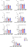
Serum concentrations of selected proteins. Serum samples were obtained after blood collection at two time points post-infection, week 6 (control and anti-TNF groups) and week 9 (control+Sb and anti-TNF+Sb groups). Seven proteins were evaluated by sandwich ELISA: (A) Fibronectin, (B) Vitronectin, (C) Transferrin, (D) Hemopexin, (E) Caveolin, (F) Dipeptidyl pptidase-4 and (G) High mobility group box 1. The concentration of each selected protein is shown individually. Also provided are the means and standard errors for each study group. Comparisons were made between groups sharing the same immunosuppression status (control vs. control + Sb and anti-TNF vs. anti-TNF + Sb), as well as between groups before (control vs. anti-TNF) and after (control + Sb vs. anti-TNF + Sb) VL treatment. Significant differences were assessed using an ANOVA followed by a Tukey’s post hoc test and are indicated as *p ≤ 0.05; **p ≤ 0.01.

Similar articles

References

    1. Gradoni L, Lopez-Velez R, Mokni M. Manual on case management and surveillance of the leishmaniases in the WHO European Region. Geneva, Switzerland: World Health Organisation; (2017).
    1. Kurizky PS, Marianelli FF, Cesetti MV, Damiani G, Sampaio RNR, Goncalves LMT, et al. A comprehensive systematic review of leishmaniasis in patients undergoing drug-induced immunosuppression for the treatment of dermatological, rheumatological and gastroenterological diseases. Rev Inst Med Trop Sao Paulo. (2020) 62:e28. doi: 10.1590/s1678-9946202062028, PMID: - DOI - PMC - PubMed
    1. Bosch-Nicolau P, Ubals M, Salvador F, Sanchez-Montalva A, Aparicio G, Erra A, et al. Leishmaniasis and tumor necrosis factor alpha antagonists in the Mediterranean basin. A switch in clinical expression. PloS Negl Trop Dis. (2019) 13:e0007708. doi: 10.1371/journal.pntd.0007708, PMID: - DOI - PMC - PubMed
    1. Neumayr AL, Morizot G, Visser LG, Lockwood DN, Beck BR, Schneider S, et al. Clinical aspects and management of cutaneous leishmaniasis in rheumatoid patients treated with TNF-alpha antagonists. Travel Med Infect Dis. (2013) 11:412–20. doi: 10.1016/j.tmaid.2013.05.003, PMID: - DOI - PubMed
    1. van Griensven J, Carrillo E, López-Vélez R, Lynen L, Moreno J. Leishmaniasis in immunosuppressed individuals. Clin Microbiol Infection. (2014) 20:286–99. doi: 10.1111/1469-0691.12556, PMID: - DOI - PubMed

MeSH terms

LinkOut - more resources